
Diagnosis confirmed by EGD findings and clinical presentation
Study method
Ultrasound
Clinical information
A 78-year-old female patient presented with a 24-hour history of epigastric pain, melena, and recurrent episodes of vomiting. During the last episodes of vomiting, streaks of blood appeared in the vomitus.
The patient was admitted to the emergency department by ambulance for further evaluation.
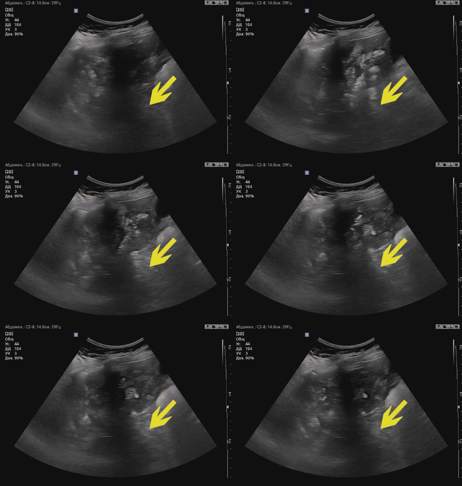
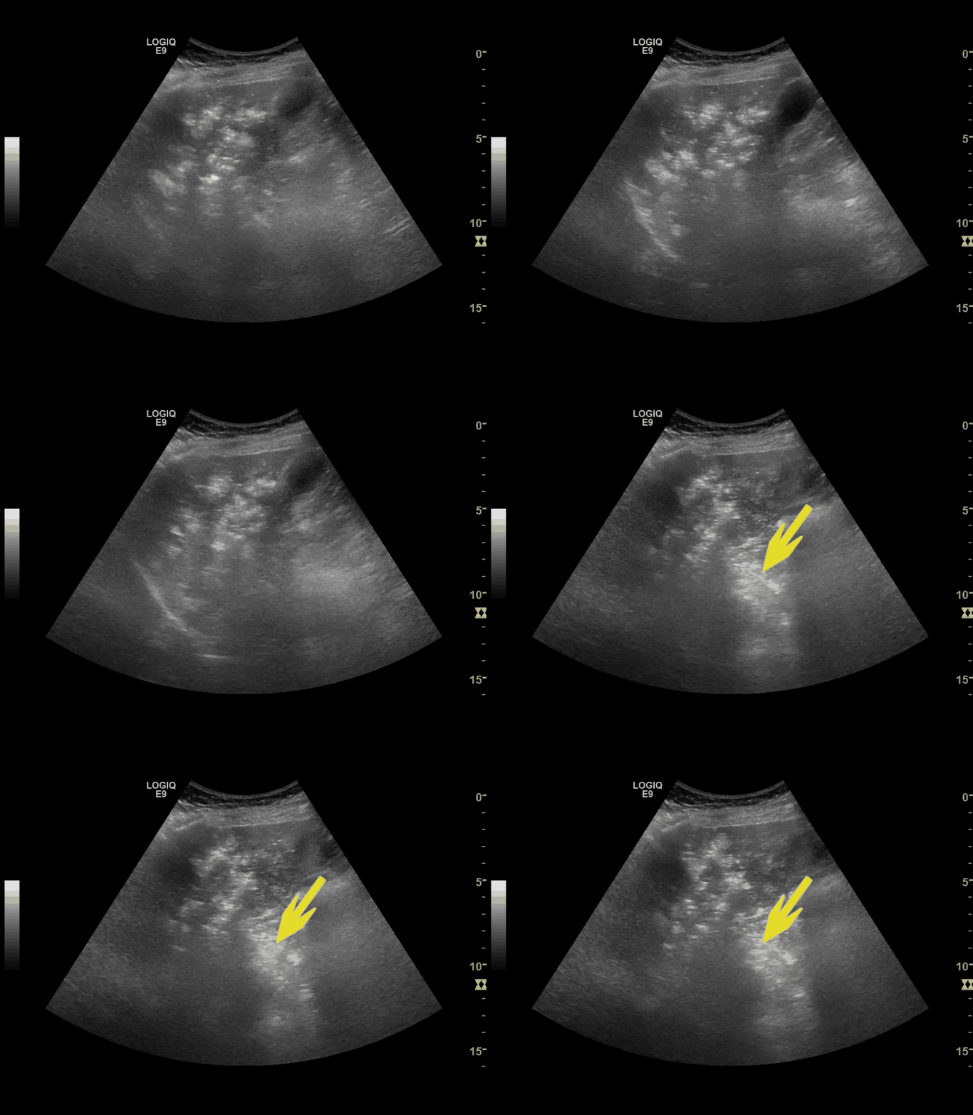
Description of sonographic findings
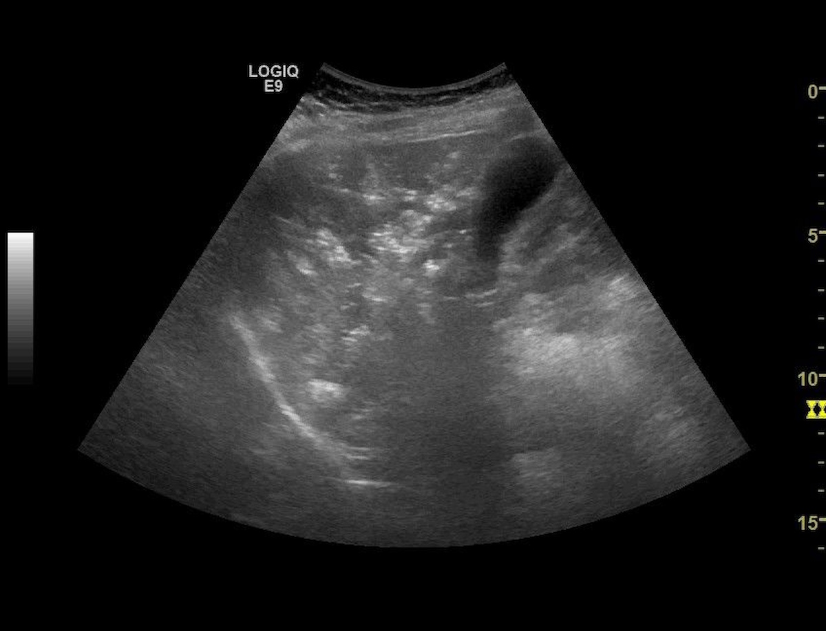
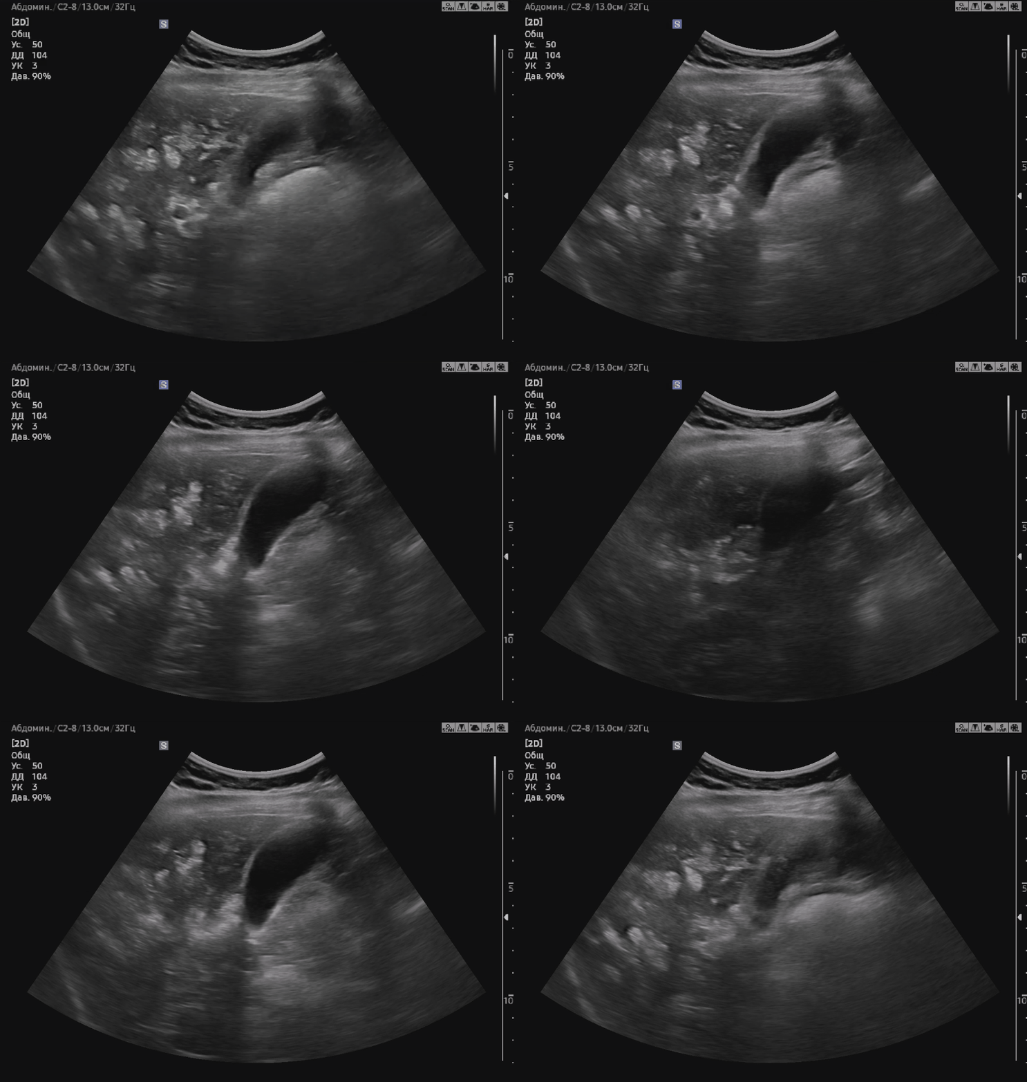
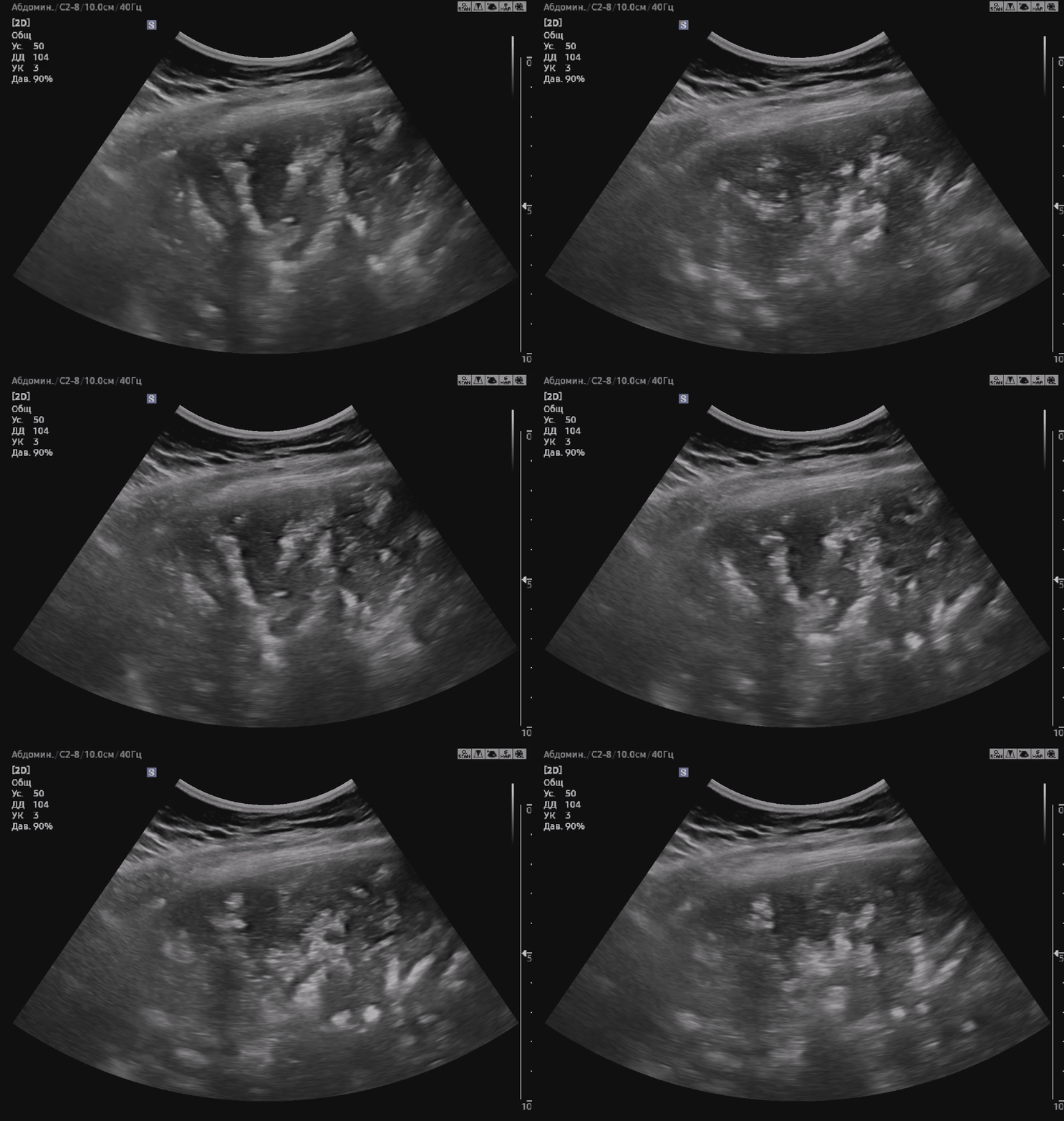
The liver was partially assessed through intercostal approaches. The hepatic parenchyma demonstrated markedly heterogeneous echotexture due to multiple branching hyperechoic foci forming a pattern resembling a vascular tree. Multiple small mobile hyperechoic foci were visualized within the lumen of the portal vein.
Conclusion of sonographic findings
The ultrasound findings are most consistent with gas within the portal vein and intrahepatic portal venous branches (hepatic portal venous gas). The differential diagnosis includes spontaneous echogenicity (spontaneous echo contrast).
Pitfall
Mobile hyperechoic echoes within the portal vein may mimic spontaneous echo contrast. However, the presence of multiple branching hyperechoic structures within the liver parenchyma, forming a vascular-tree pattern, together with the characteristic clinical presentation, strongly supports the diagnosis of hepatic portal venous gas.
Treatment
Esophagogastroduodenoscopy (EGD)
Two linear mucosal tears measuring up to 1.2 cm were identified in the gastric cardia region. The tears contained thrombosed vessels at the base and signs of previous bleeding, consistent with Mallory–Weiss syndrome. A combined endoscopic hemostatic procedure was performed: injection therapy with adrenaline solution; endoscopic clipping of bleeding vessels (5 hemostatic clips applied).
Final endoscopic diagnosis: Endoscopic findings consistent with Mallory–Weiss syndrome. Status post successful endoscopic hemostasis.
Final diagnosis
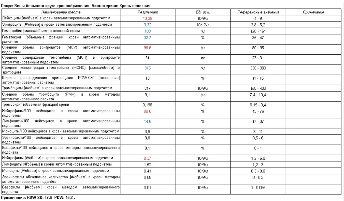
Mallory–Weiss syndrome complicated by upper gastrointestinal bleeding. Status post successful combined endoscopic hemostasis. Mild secondary posthemorrhagic iron-deficiency anemia (Hb 103 g/L).

Author
Egor Isaev
Ultrasound physician and radiologist.
Founder of an educational project featuring clinical cases in ultrasound and radiology.