
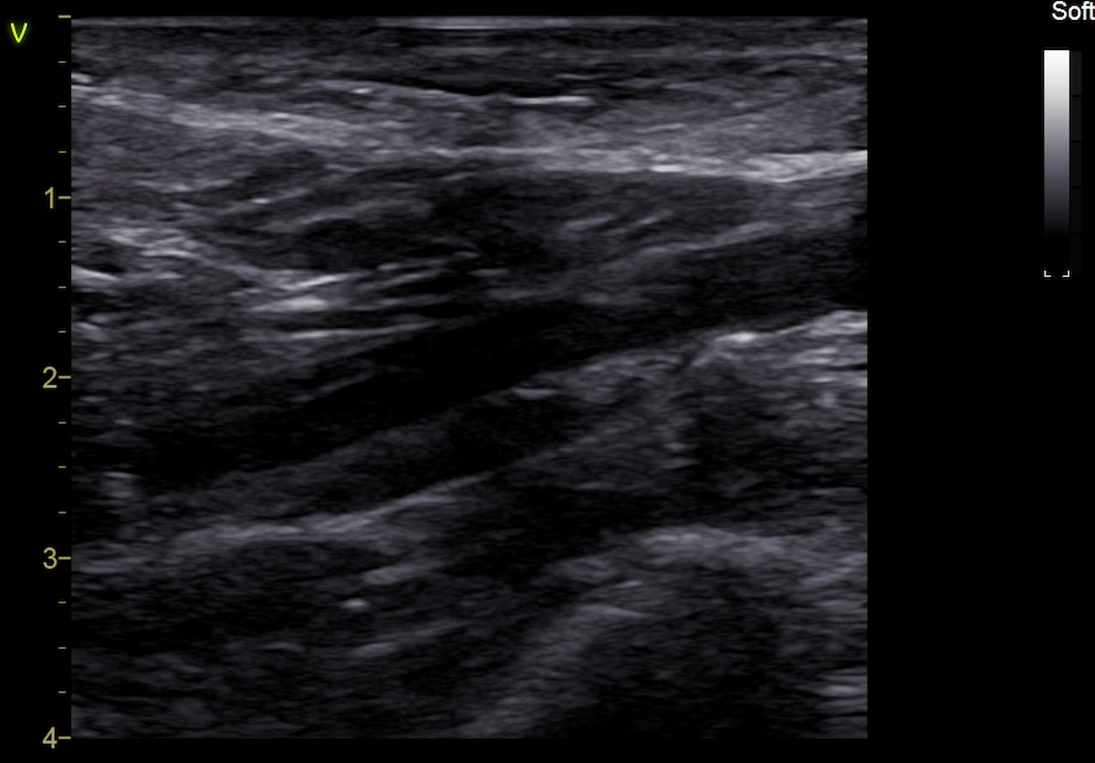
The diagnosis was supported by concordant ultrasound and MRI findings.
MRI confirmed an intramural hematoma of the right internal carotid artery.
Study method
Ultrasound, CT, MRI
Clinical information
The patient presented with dizziness, neck pain radiating to the right arm, and numbness involving the right side of the face and right arm. The symptoms had persisted for approximately one month.
During outpatient evaluation, MR angiography demonstrated abnormalities of the right internal carotid artery requiring differential diagnosis between arterial dissection and aneurysmal dilatation. The patient was subsequently admitted to the hospital for further evaluation and diagnostic clarification.


Description of sonographic findings
In the midportion of the extracranial right internal carotid artery, eccentric hypoechoic wall thickening measuring up to 2.5–2.6 mm is visualized, corresponding to an intramural hematoma.
The outer diameter of the artery increases from 4.1 to 6.7 mm in the affected segment, while the lumen remains nearly unchanged, decreasing only from 4.1 to 3.9 mm. No internal blood flow is detected within the intramural hematoma on color Doppler imaging.
No focal increase in flow velocities or hemodynamically significant stenosis of the right ICA is identified.
Conclusion of sonographic findings
Ultrasound findings consistent with right internal carotid artery dissection with an intramural hematoma and focal increase in the outer arterial diameter, without hemodynamically significant stenosis.

Description of radiographic findings
CT angiography demonstrates opacification of the common, external, and internal carotid arteries. No hemodynamically significant stenosis is identified.
Conclusion of radiographic findings
No convincing CT angiographic evidence of carotid artery dissection.


T2 TSE
Description of MR findings
An intramural hematoma is identified within the wall of the right internal carotid artery, extending approximately 34 mm from the ICA origin to the C2 level and measuring up to 3 mm in thickness. The ICA lumen at this level measures up to 4.3 mm.
Conclusion of MR findings
MRI findings consistent with an intramural hematoma of the right internal carotid artery.
Final diagnosis
Right internal carotid artery dissection with intramural hematoma.
Pitfall
The absence of arterial luminal stenosis does not exclude dissection.
In this case, the intramural hematoma predominantly resulted in an increase in the outer diameter of the right ICA from 4.1 to 6.7 mm, while the lumen remained nearly unchanged, decreasing only from 4.1 to 3.9 mm. No hemodynamically significant stenosis or focal increase in flow velocity was identified.
The arterial wall abnormality was clearly visualized on ultrasound, whereas subsequent CT angiography showed no convincing evidence of dissection. Persistent clinical and imaging suspicion prompted further evaluation, and MRI subsequently confirmed an intramural hematoma of the right ICA.
Key point: in arterial dissection, an intramural hematoma may expand predominantly toward the outer contour of the vessel without causing substantial luminal narrowing. Therefore, preservation of the arterial lumen and the absence of significant stenosis do not exclude dissection. When imaging findings are discordant and clinical suspicion persists, assessment should extend beyond the arterial lumen to include the vessel wall, with additional imaging performed when appropriate.
