
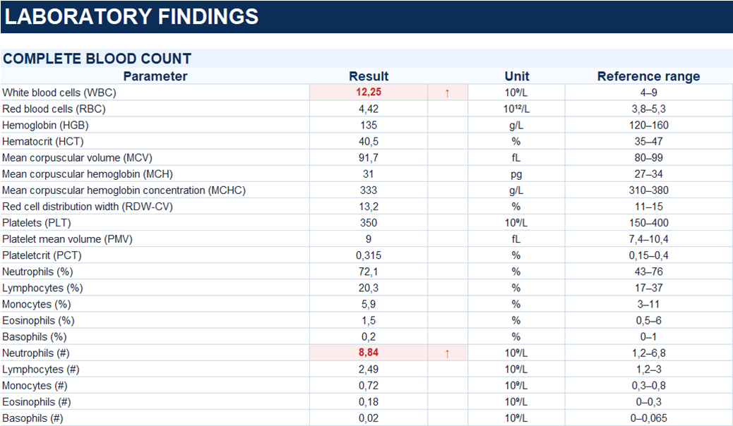
The diagnosis was established based on the clinical presentation and concordant ultrasound and contrast-enhanced CT findings.
Study method
Ultrasound, CT
Clinical information
A 51-year-old woman was admitted with lower abdominal pain, predominantly in the left iliac fossa, persisting for approximately 24 hours.
Her medical history included colonic resection with temporary colostomy for perforated diverticulitis nine years earlier. Intestinal continuity was subsequently restored.
Description of sonographic findings
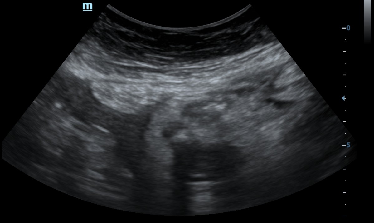
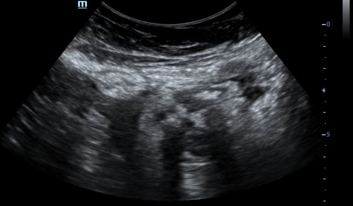
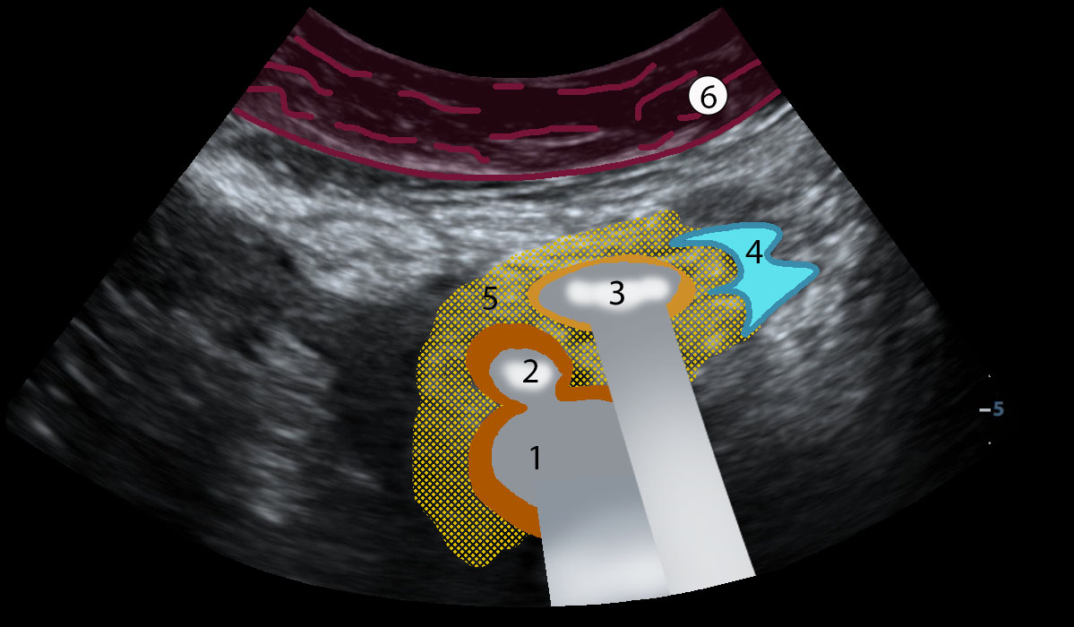
In the left flank, a focally abnormal segment of the colon is identified, with wall thickening up to 5 mm. Normal bowel wall stratification is preserved.
An 11 × 11 mm diverticulum is visualized, containing hyperechoic material with posterior acoustic shadowing, likely due to gas and/or dense fecal material.
The surrounding pericolic fat is hyperechoic, with scattered thin fluid layers measuring up to 4–5 mm, without evidence of a well-defined fluid collection.
Conclusion of sonographic findings
Sonographic findings of acute diverticulitis with localized inflammatory changes in the surrounding pericolic fat.
Contrast-enhanced CT, portal venous phase.
Description of radiographic findings
Contrast-enhanced abdominal CT demonstrates multiple diverticula in the descending and sigmoid colon.
At the junction of the descending and sigmoid colon, focal bowel wall thickening and inflammatory stranding of the surrounding pericolic fat are present.
Additionally, two ventral hernias of the linea alba are identified in the mid-abdomen. The superior hernia contains a segment of the transverse colon, while the inferior hernia contains small bowel loops.
No free intraperitoneal fluid is identified.
Conclusion of radiographic findings
CT findings of acute diverticulitis at the descending–sigmoid colon junction (modified Hinchey Ia).
Additionally, two ventral hernias of the linea alba.
Treatment
Conservative treatment was administered, including antibiotic therapy (ciprofloxacin and metronidazole) and symptomatic management.
Final diagnosis
Colonic diverticular disease. Acute diverticulitis at the descending–sigmoid colon junction (modified Hinchey Ia).
Key message
Ultrasound identified an inflamed diverticulum, focal bowel wall thickening, and inflammatory changes in the surrounding pericolic fat.
CT confirmed inflammatory changes at the junction of the descending and sigmoid colon.
A history of perforated diverticulitis requiring colonic resection is an important diagnostic clue when evaluating a patient presenting with recurrent abdominal pain