
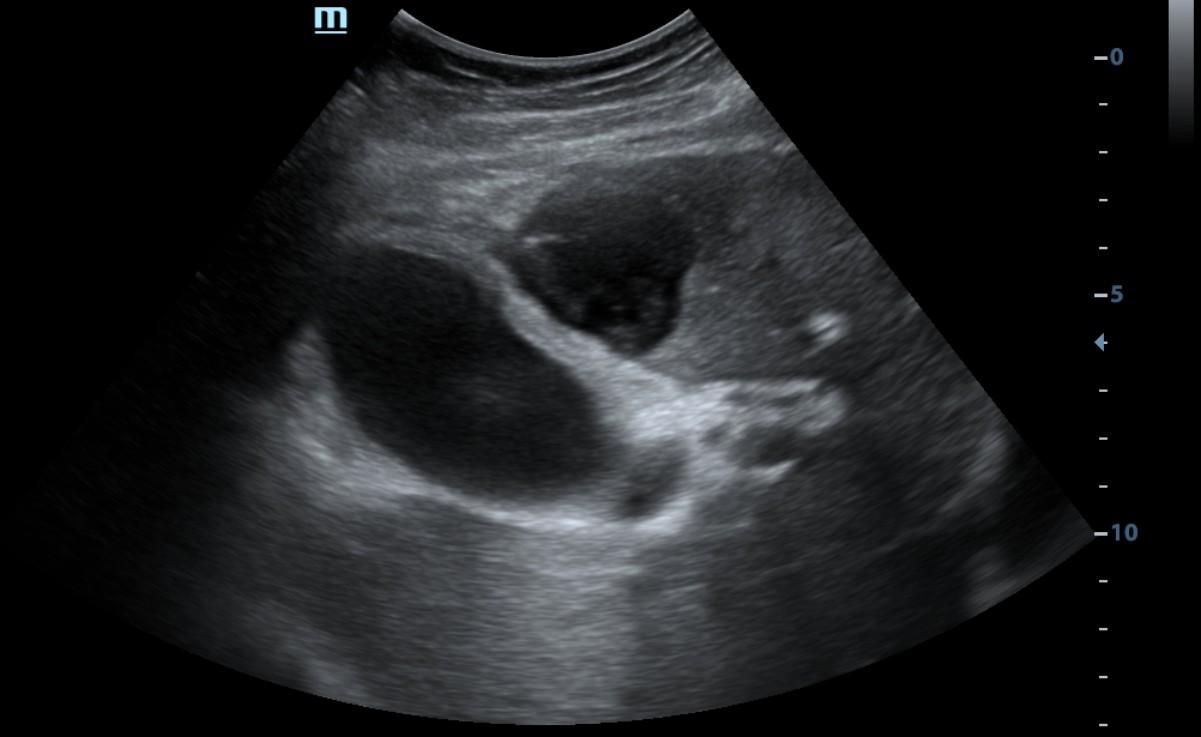
The diagnosis was established based on concordant ultrasound and CT findings. Acute calculous cholecystitis was additionally confirmed by findings obtained during percutaneous cholecystostomy.
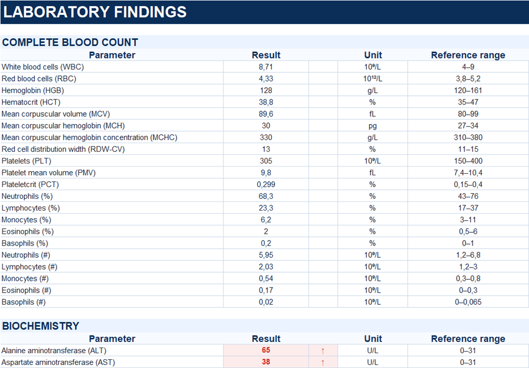
Study method
Ultrasound, CT
Clinical information
A 69-year-old woman initially presented with repeated vomiting, followed by pain in the right upper quadrant and epigastric region. She was admitted to the hospital due to these symptoms.
Description of sonographic findings
The gallbladder is distended, measuring 87 × 51 mm, and is noncompressible. Its wall is thickened to 4 mm and demonstrates focal stratification.
The cystic duct is dilated to 11 mm, with an impacted calculus measuring up to 12 mm in its lumen.
An irregular, heterogeneous fluid collection measuring 39 × 36 mm is identified in the adjacent hepatic parenchyma, in the region of segment IV.
The surrounding pericholecystic fat is edematous, with thin fluid layers measuring up to 4 mm.
The intrahepatic and extrahepatic bile ducts are not dilated. The common bile duct measures 5 mm.
A small amount of free fluid, measuring up to 3 mm in thickness, is present in the subhepatic space.
Conclusion of sonographic findings
Sonographic findings of acute calculous cholecystitis with marked pericholecystic inflammatory changes and an adjacent hepatic fluid collection suspicious for abscess formation.
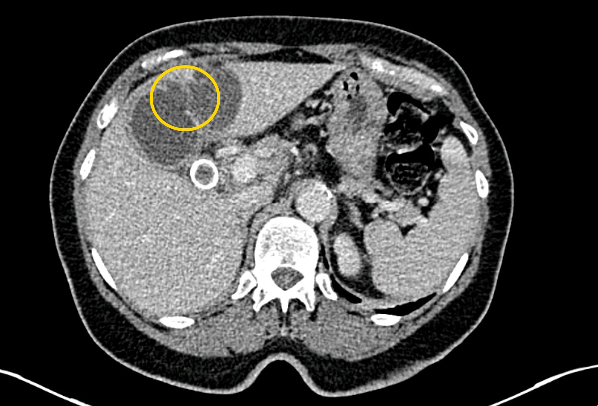
Contrast-enhanced CT, portal venous phase.
Description of radiographic findings
On contrast-enhanced abdominal CT, the gallbladder is distended, with a radiopaque calculus measuring up to 21 mm in the gallbladder neck.
The gallbladder wall is thickened. A focal wall defect measuring approximately 14 mm is identified along the anterior aspect of the gallbladder body, communicating with an adjacent subcapsular hepatic fluid collection measuring up to 43 × 35 mm.
The surrounding fat demonstrates inflammatory stranding.
The intrahepatic and extrahepatic bile ducts are not dilated.
Conclusion of radiographic findings
CT findings of acute calculous cholecystitis complicated by localized gallbladder perforation with formation of an adjacent subcapsular hepatic abscess.
Treatment
Ultrasound-guided percutaneous transhepatic cholecystostomy was performed, with drainage of 50 mL of turbid bile.
Follow-up fistulography confirmed correct catheter positioning, with no contrast leakage detected.
The patient was discharged with a recommendation for subsequent consideration of elective laparoscopic cholecystectomy.
Final diagnosis
Acute calculous cholecystitis with localized gallbladder wall perforation and formation of an adjacent subcapsular hepatic abscess (Niemeier type II, based on imaging findings).
Pitfall
In localized gallbladder perforation, the wall defect may not be directly visualized on ultrasound. An adjacent fluid collection may therefore be mistaken for a primary hepatic abscess unrelated to gallbladder disease.
The presence of a fluid collection near an inflamed gallbladder should raise suspicion for perforation, even when no definite wall defect is identified.
Key message
Ultrasound demonstrated acute calculous cholecystitis with an adjacent fluid collection suspicious for abscess formation.
Contrast-enhanced CT identified a focal defect in the gallbladder wall communicating with the adjacent subcapsular hepatic fluid collection, characteristic of localized gallbladder perforation (Niemeier type II).