
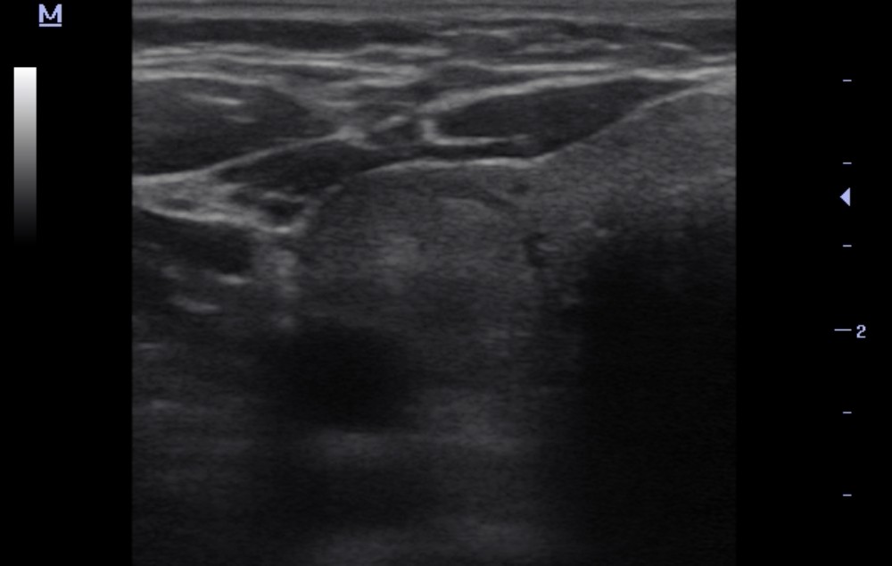
The cytologic diagnosis was confirmed by fine-needle aspiration (FNA).
Cytologic findings are consistent with a follicular thyroid neoplasm (Bethesda IV).
Study method
Ultrasound
Clinical information
A 64-year-old woman was referred for thyroid ultrasound.
Description of sonographic findings
Isthmus: not thickened, with preserved echotexture.
Right lobe:
Not enlarged.
Contours are mildly irregular and well defined.
The parenchyma is focally heterogeneous due to the following nodules:
– Upper third: an 8.2 × 6.1 mm nodule. The nodule has a spongiform appearance. Margins are mildly irregular but well defined. A thin, subtle hypoechoic halo is present. Small hyperechoic foci are seen. Color Doppler demonstrates predominantly perinodular vascularity with occasional intranodular vascular signals. EU-TIRADS 2.
– Middle third: an 18 mm round nodule. The nodule is solid, moderately hyperechoic, and mildly heterogeneous. Margins are smooth and well defined. A hypoechoic halo is present. Color Doppler demonstrates prominent predominantly perinodular vascularity with occasional intranodular vascular signals. EU-TIRADS 3.
– Lower third, adjacent to the isthmus: a 9.1 × 4.8 mm nodule. The nodule has a spongiform appearance. Margins are mildly irregular but well defined. A thin, subtle hypoechoic halo is present. Small hyperechoic foci are seen. Color Doppler demonstrates predominantly perinodular vascularity with occasional intranodular vascular signals. EU-TIRADS 2.
No increased vascularity of the thyroid parenchyma is seen on color or power Doppler imaging.
Left lobe:
Not enlarged.
Contours are irregular and well defined.
The parenchyma is focally heterogeneous due to the following nodules:
– Two spongiform nodules measuring 4.4 mm and 7.7 mm, respectively, are seen in the upper and middle thirds of the lobe. Their margins are mildly irregular but well defined. Color Doppler demonstrates predominantly perinodular vascularity with occasional intranodular vascular signals. EU-TIRADS 2.
No increased vascularity of the thyroid parenchyma is seen on color or power Doppler imaging.
Conclusion of sonographic findings
Sonographic findings of multiple bilateral thyroid nodules; EU-TIRADS categories are provided in the description.
Verification Method
Fine-needle aspiration (FNA) of nodules in both thyroid lobes was performed, followed by cytologic examination.
Right thyroid nodule:
The cytologic specimen contains solid and follicle-like groups of thyroid follicular cells with round, hyperchromatic, monomorphic nuclei and moderately prominent nucleoli. Some cell clusters show foci of residual colloid formation.
Cytologic diagnosis: follicular thyroid neoplasm — Bethesda IV.
Left thyroid nodule:
The cytologic specimen contains abundant homogeneous colloid, occasional macrophages, and small groups and isolated thyroid follicular cells without cytologic atypia.
Cytologic diagnosis: colloid nodule — Bethesda II.
Final diagnosis
Follicular neoplasm of the right thyroid lobe (Bethesda IV). Colloid nodule of the left thyroid lobe (Bethesda II).
Key message
Ultrasound: 18 mm solid right thyroid nodule — EU-TIRADS 3 → FNA: Bethesda IV.
Ultrasound: spongiform left thyroid nodule — EU-TIRADS 2 → FNA: Bethesda II.
EU-TIRADS estimates the sonographic risk of malignancy, whereas Bethesda reflects the results of cytologic assessment; there is no direct correspondence between the categories of these two systems.
Bethesda IV does not distinguish follicular adenoma from follicular carcinoma, as this requires histologic assessment of capsular and vascular invasion.
The 18 mm right thyroid nodule is below the standard FNA size threshold for EU-TIRADS 3 (>20 mm); no additional indication for biopsy was documented in the available medical records.