
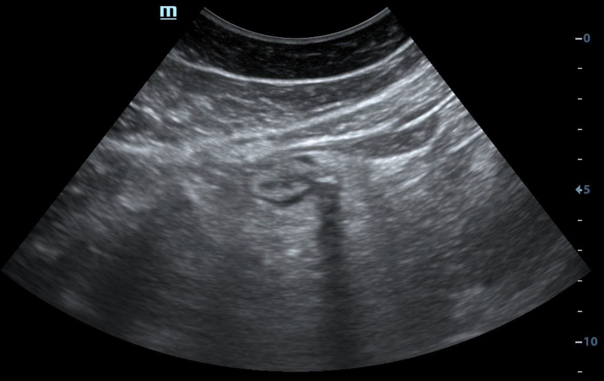
The diagnosis was confirmed by concordant ultrasound and CT findings
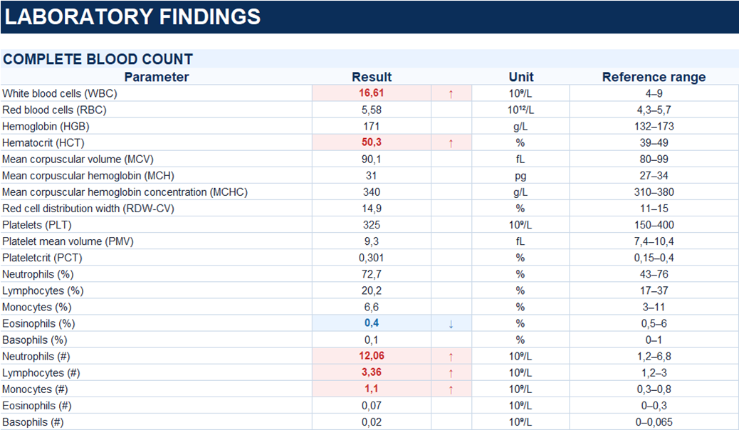
Study method
Ultrasound, CT
Clinical information
A 54-year-old male patient presented with pain in the left flank, abdominal bloating, and fever up to 38 °C. He denied nausea and vomiting. He had taken NSAIDs without improvement. Due to persistent symptoms, he was admitted to the hospital on an emergency basis.

Description of sonographic findings
In the left iliac region, a colonic segment is visualized with a diverticular outpouching measuring up to 10.7 × 11.4 mm and a neck approximately 6 mm wide. Within the diverticular lumen, a hyperechoic component with posterior acoustic shadowing is identified, likely due to gas and/or dense fecal material. The wall of the adjacent colonic segment is thickened to 5 mm, predominantly due to thickening of the submucosal layer. The surrounding pericolic fat demonstrates edema and inflammatory infiltration.
Conclusion of sonographic findings
Sonographic findings of acute diverticulitis with surrounding inflammatory changes in the pericolic fat.

Contrast-enhanced CT, portal venous phase.
Description of radiographic findings
At the junction of the descending and sigmoid colon, there is a diverticular outpouching measuring up to 12 × 12 mm, with a thickened, partially calcified wall measuring up to 3 mm. The adjacent bowel wall is circumferentially thickened up to 7 mm. Inflammatory changes are present in the surrounding pericolic fat.
A small amount of fluid, measuring up to 5 mm in thickness, is present along the left retrocolic fascia, with inflammatory changes in the adjacent fat. A small amount of free fluid is also present in the pelvis.
Conclusion of radiographic findings
CT findings of acute diverticulitis at the descending–sigmoid colon junction with surrounding inflammatory changes (modified Hinchey Ia). A small amount of free fluid is present in the abdominal and pelvic cavities.
Final diagnosis
Colonic diverticular disease. Acute diverticulitis at the descending–sigmoid colon junction (modified Hinchey Ia), without evidence of free perforation or abscess formation.