

The diagnosis was confirmed by concordant clinical and ultrasound findings.
The diagnosis was confirmed intraoperatively.
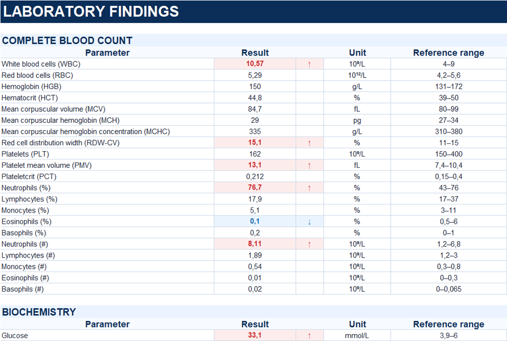
Marked hyperglycemia was also present (serum glucose, 33.1 mmol/L) in the setting of type 2 diabetes mellitus.
Study method
Ultrasound
Clinical information
A 55-year-old patient presented with a two-day history of pain in the perianal region. He denied nausea, vomiting, and changes in bowel habits. Due to persistent symptoms, he was admitted to the hospital.
On physical examination, a hyperemic and edematous mass measuring approximately 2 × 3 cm was identified in the perianal region at the 2 o’clock position and was markedly tender to palpation.

Description of sonographic findings
In the soft tissues of the perianal region, along the medial aspect of the left buttock at the 7 o’clock position, there is a heterogeneous fluid collection measuring up to 29 × 19 mm, located approximately 5 mm beneath the skin surface. The surrounding fat demonstrates increased echogenicity. Multiple foci of increased vascularity are present along the periphery of the collection.
Conclusion of sonographic findings
Sonographic findings of a perianal soft-tissue abscess with surrounding inflammatory changes.
Treatment
Surgical incision and drainage of the perianal abscess were performed.
Final diagnosis
Acute subcutaneous paraproctitis with perianal abscess formation.