
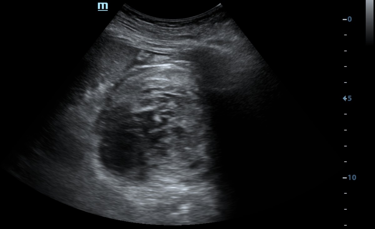
The diagnosis was established based on the clinical history and concordant ultrasound and contrast-enhanced CT findings.
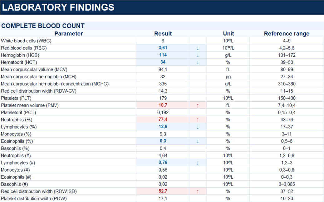
Study method
Ultrasound, CT
Clinical information
A 63-year-old male patient, G., was admitted after falling into a construction pit from standing height and striking the left side of his chest against a metal pipe.
He complained of left-sided chest pain aggravated by inspiration, dyspnea, and right knee pain.
Green arrow — lower pole of the left kidney; yellow arrow — psoas major muscle; blue arrow — heterogeneous fluid collection (hematoma); red arrow — iliac vessels.
Yellow arrow — psoas major muscle; blue arrow — heterogeneous fluid collection (hematoma); red arrow — iliac vessels.
Description of sonographic findings
Multiple cystic lesions are identified in both kidneys.
In the middle third of the left kidney, an 83 × 61 mm cystic lesion containing echogenic material is visualized. An additional lesion of similar echotexture, measuring 33 × 28 mm, is located adjacent to it.
The surrounding perinephric fat is heterogeneous and infiltrated, with a small amount of fluid.
In the left retroperitoneal space, extending from the level of the lower pole of the spleen toward the pelvis, a fluid collection interpreted as a hematoma is identified, with a maximum thickness of 27 mm.
A small amount of free anechoic fluid, estimated at less than 100 mL, is present in the peritoneal cavity.
Conclusion of sonographic findings
Sonographic findings of hemorrhagic changes involving a cystic lesion of the left kidney (hemorrhage into a cyst? Adjacent hematoma?). Left retroperitoneal hematoma. Small amount of free intraperitoneal fluid.
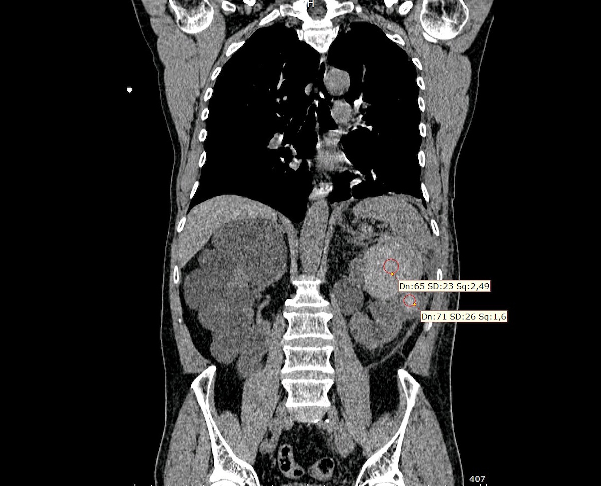
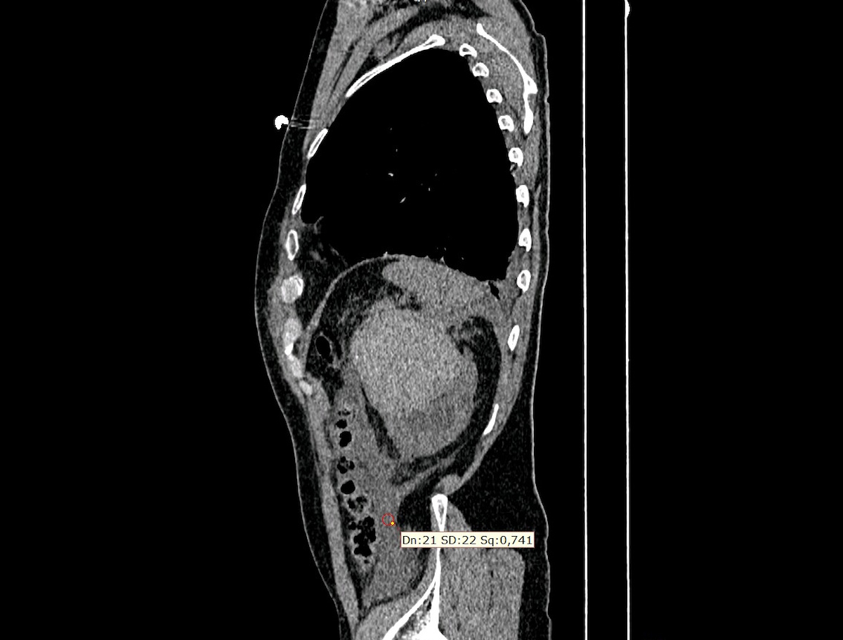
Multiphase CT. Arterial and portal venous phases.
Description of radiographic findings
Contrast-enhanced CT of the chest and abdomen demonstrates multiple cysts of homogeneous fluid attenuation in both kidneys, measuring up to 111 × 86 mm.
In the region of the upper pole of the left kidney, predominantly superior and anterior to it, a heterogeneous lesion measuring 68 × 95 × 82 mm is identified. It has relatively well-defined margins and areas of increased attenuation (+65–71 HU on noncontrast images), consistent with hemorrhagic contents.
Hemorrhagic changes are identified along the periphery of the left kidney. The left perinephric fat demonstrates stranding and infiltration.
An extensive fluid collection of relatively low attenuation (+17–21 HU), presumed to be post-traumatic, is visualized in the left retroperitoneal space along the renal fascial planes.
No contrast extravasation is identified.
The renal collecting systems are not dilated. Contrast excretion by both kidneys is timely and symmetric.
Additional findings include fractures of the left ninth through eleventh ribs and a small amount of fluid in the left pleural cavity.
Conclusion of radiographic findings
CT findings of traumatic hemorrhage into a left renal cyst with adjacent perirenal hemorrhagic changes and an extensive left retroperitoneal fluid collection, likely post-traumatic in nature. No evidence of active contrast extravasation.
Associated findings: fractures of the left ninth through eleventh ribs, small left-sided pleural effusion, and multiple bilateral renal cysts.
Treatment
No signs of active contrast extravasation were identified on CT; therefore, no indications for emergency surgical intervention were established at the time of assessment. Conservative management was chosen, with serial ultrasound examinations and hemoglobin monitoring.
Follow-up ultrasound examinations demonstrated no significant changes in the left renal findings. The maximum thickness of the retroperitoneal fluid collection decreased from 27 to 8 mm.
Final diagnosis
Combined blunt trauma to the chest and abdomen. Blunt injury to the left kidney with hemorrhage into a cyst, formation of a perirenal hematoma, and an associated left retroperitoneal fluid collection. Multiple bilateral renal cysts. Fractures of the left ninth through eleventh ribs.
Key message
Ultrasound revealed heterogeneous contents within cystic lesions of the left kidney and an extensive retroperitoneal fluid collection, raising suspicion of traumatic hemorrhage.
Contrast-enhanced CT demonstrated high-attenuation hemorrhagic contents in the region of the left kidney; no evidence of active contrast extravasation was identified.
Despite the relatively low attenuation of the retroperitoneal fluid collection (+17–21 HU), its hemorrhagic nature is considered likely in the context of trauma, concomitant hemorrhage in the region of the left kidney, and findings from follow-up examinations. However, low fluid attenuation alone can neither confirm nor exclude the presence of blood.
