
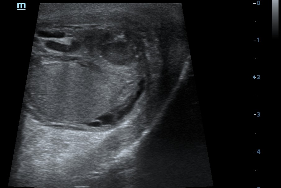
The diagnosis was established based on the clinical presentation and ultrasound findings.
The abscess of the left epididymal tail was confirmed intraoperatively during scrotal exploration.
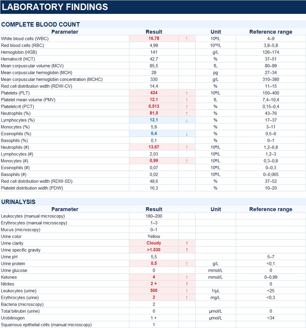
Study method
Ultrasound
Clinical information
A 77-year-old male patient, R., was admitted with complaints of bilateral scrotal pain and swelling, accompanied by fever up to 39 °C.
The patient had undergone a transrectal prostate biopsy, followed by the onset of fever later that same day. Scrotal pain and swelling developed several days afterward. Levofloxacin was prescribed on an outpatient basis. Due to persistent symptoms, the patient was hospitalized.
Description of sonographic findings
Right testis: measures 45 × 27 × 28 mm, with preserved echotexture and no focal lesions.
The right epididymis is enlarged, with the head measuring up to 12 mm and the tail up to 23 mm. The echotexture is diffusely heterogeneous.
Left testis: measures 44 × 29 × 23 mm, with preserved echotexture and no focal lesions.
The left epididymis is enlarged, with the head measuring up to 12 mm and the body up to 16 mm. The echotexture is diffusely heterogeneous.
In the region of the left epididymal tail, a heterogeneous hypoechoic lesion measuring 11 × 10 mm is identified, with a poorly delineated wall, posterior acoustic enhancement, and hyperechoic foci (possibly representing gas). Power Doppler imaging demonstrates no internal vascularity, with marked peripheral hypervascularity. The sonographic appearance is consistent with an abscess.
Free fluid: Fluid is present between the layers of the tunica vaginalis, measuring up to 11 mm in thickness on the right and up to 6 mm on the left. The fluid on the left is heterogeneous, with fibrin strands.
Conclusion of sonographic findings
Sonographic findings of acute bilateral epididymitis with abscess formation in the left epididymal tail. Free fluid within the scrotal cavity bilaterally, heterogeneous on the left.
Treatment
Surgical exploration of the left hemiscrotum was performed. The epididymis was enlarged and indurated, with an area of fluctuation in the tail. Upon incision of the abscess, 15 mL of purulent material was drained. Drainage of the left hemiscrotum was performed.
Antibiotic therapy with ceftriaxone was prescribed.
Final diagnosis
Acute bilateral epididymitis. Abscess of the left epididymal tail.
Key message
An epididymal abscess may appear as a heterogeneous hypoechoic lesion with a surrounding wall and posterior acoustic enhancement. Doppler ultrasound typically demonstrates absent internal vascularity in combination with peripheral hypervascularity.
In this case, the combination of these sonographic findings in the setting of acute epididymitis raised suspicion of an abscess in the left epididymal tail, which was subsequently confirmed intraoperatively.