
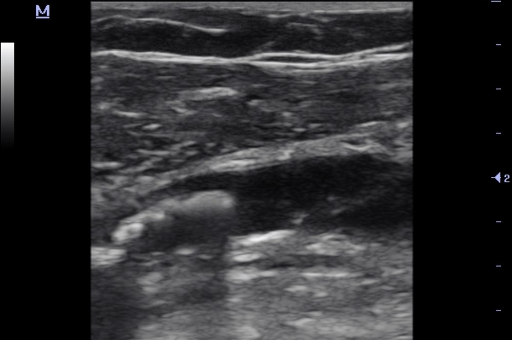
Outpatient duplex ultrasonography revealed an atherosclerotic plaque in the left common femoral artery with a mobile (floating) component.
Subsequent surgical intervention in the same artery confirmed the presence of a stenosing atherosclerotic plaque with an unstable component.
Study method
Ultrasound
Clinical information
A 67-year-old female patient, I., attended an outpatient clinic for a scheduled duplex ultrasound examination of the lower extremity veins to exclude thrombosis before an upcoming surgical procedure unrelated to vascular disease. At the time of examination, she reported no lower extremity symptoms or general complaints.
During the ultrasound examination, an atherosclerotic plaque with a mobile (floating) component was additionally identified in the left common femoral artery.
Due to this finding, emergency medical services were called, and the patient was transported to a hospital for further evaluation and treatment.
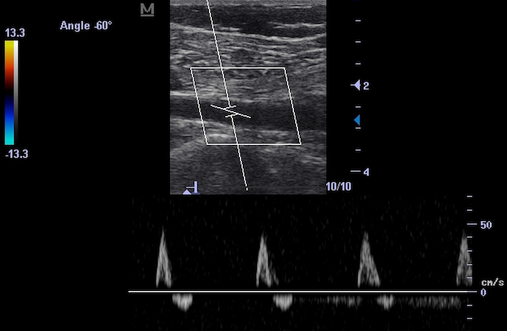
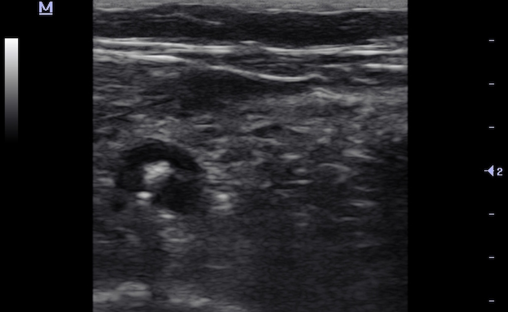
Description of sonographic findings
A heterogeneous atherosclerotic plaque with areas of calcification is identified in the left common femoral artery, causing up to 65% luminal narrowing by diameter.
The plaque demonstrates features of instability: a mobile (floating) component measuring up to 13 mm in length is visualized.
Distal to the stenosis, a predominantly normal arterial flow waveform is recorded.
Conclusion of sonographic findings
Sonographic findings of atherosclerotic disease of the left common femoral artery, with up to 65% stenosis by diameter and features of plaque instability (a floating component, presumably thrombotic in nature).
Treatment
Catheter angiography was performed, revealing up to 70% stenosis of the left common femoral artery.
During subsequent surgical intervention, an atherosclerotic plaque with an unstable component was identified within the lumen of the left common femoral artery, causing up to 70% stenosis.
Endarterectomy with removal of the atherosclerotic plaque was performed, followed by patch angioplasty using a xenopericardial patch. Blood flow through the femoral arteries was restored.
Final diagnosis
Atherosclerosis of the lower extremity arteries. Up to 70% stenosis of the left common femoral artery caused by an atherosclerotic plaque with an unstable component.
Key message
When performing duplex ultrasonography of the lower extremity vessels, it is important to assess not only the degree of stenosis but also the morphological characteristics of atherosclerotic plaques, including the presence of mobile components.
In this case, an atherosclerotic plaque with a 13 mm floating component was incidentally identified in the left common femoral artery during a scheduled examination of the lower extremity veins. Despite the preservation of a predominantly normal arterial flow waveform distal to the stenosis, this finding prompted emergency hospitalization.
Subsequent surgical intervention in the same artery confirmed the presence of a stenosing atherosclerotic plaque with an unstable component.