
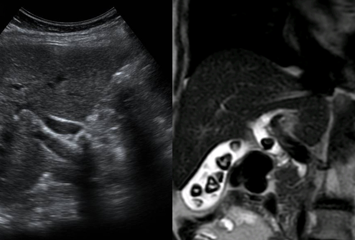
The diagnosis was established based on ultrasonography and magnetic resonance cholangiopancreatography (MRCP) findings and confirmed by endoscopic retrograde cholangiopancreatography (ERCP) with subsequent stone extraction.
Study method
Ultrasound, MRI
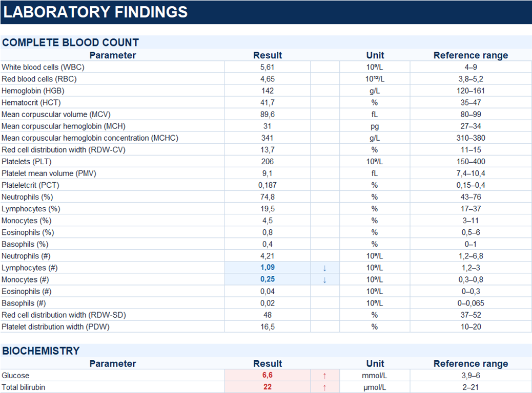
Clinical information
A 72-year-old woman was brought to the hospital by ambulance with a two-week history of epigastric and right upper quadrant pain.
Description of sonographic findings
The gallbladder measures 66 × 27 mm and is not enlarged. Multiple gallstones measuring up to 8 mm are identified within its lumen. The gallbladder wall is not thickened.
The extrahepatic bile duct is dilated to 12 mm. A stone measuring up to 12 mm is visualized within its lumen, at the level of the transition between the common hepatic duct (CHD) and the common bile duct (CBD).
Conclusion of sonographic findings
Sonographic findings of cholecystolithiasis, an extrahepatic bile duct stone at the level of the transition between the common hepatic duct and the common bile duct, and extrahepatic bile duct dilatation.
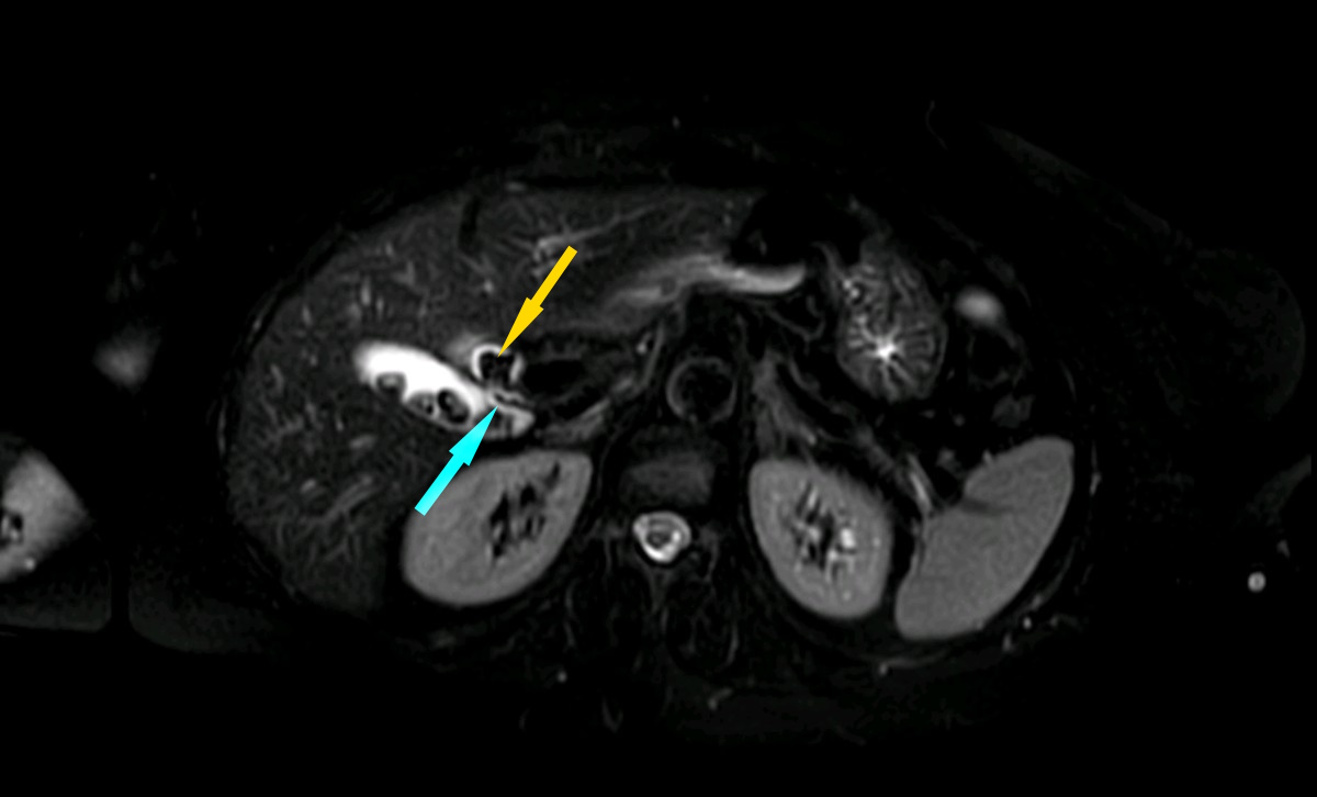
T2W TSE FS – axial
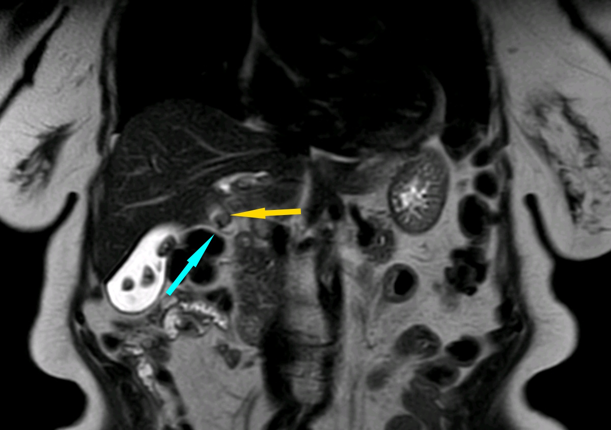
T2W TSE – coronal
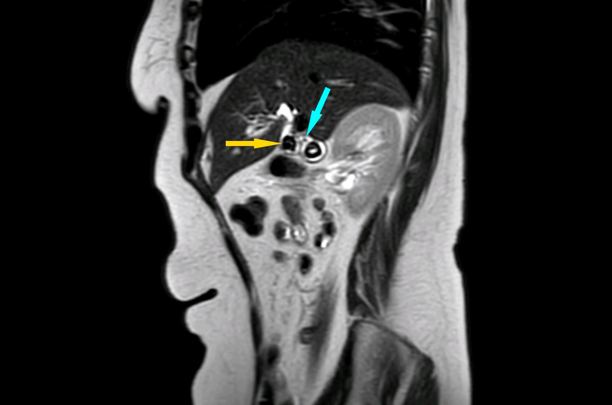
T2W TSE – sagittal
MRCP RT – coronal
Description of MR findings
The gallbladder measures 88 × 26 mm and contains multiple gallstones measuring up to 15 mm, including stones in the gallbladder neck and infundibular region. The gallbladder wall measures up to 2.5 mm in thickness, without evidence of surrounding tissue edema.
The intrahepatic bile ducts are dilated to 8 mm.
The common hepatic duct is dilated to 13 mm. A stone measuring 11 × 10 mm is identified in its distal portion, immediately proximal to the cystic duct insertion.
The common bile duct measures up to 7 mm in diameter and can be traced to the major duodenal papilla. No additional stones are convincingly identified within its lumen.
Conclusion of MR findings
MRI findings of cholecystolithiasis, a stone in the distal common hepatic duct, and biliary hypertension.
Treatment
Endoscopic retrograde cholangiopancreatography (ERCP) was performed. Cholangiography demonstrated a rounded filling defect measuring up to 10 mm within the common bile duct, consistent with a stone. The duct was dilated to 11 mm.
Endoscopic biliary sphincterotomy was performed. During subsequent bile duct exploration using a Dormia basket, the stone was fragmented and extracted.
Final diagnosis
Cholelithiasis. Cholecystolithiasis. Cholangiolithiasis.
Key message
Accurate localization of extrahepatic bile duct stones requires careful identification of the junction between the common hepatic duct and the cystic duct, which together form the common bile duct.
In this case, ultrasonography and MRCP demonstrated a stone in the region of this anatomical junction. MRCP further localized the stone to the distal common hepatic duct.
Subsequent ERCP confirmed the presence of a bile duct stone, and endoscopic stone extraction was performed. This case highlights the importance of correlating findings from different imaging modalities while considering the anatomy of the biliary tree.