
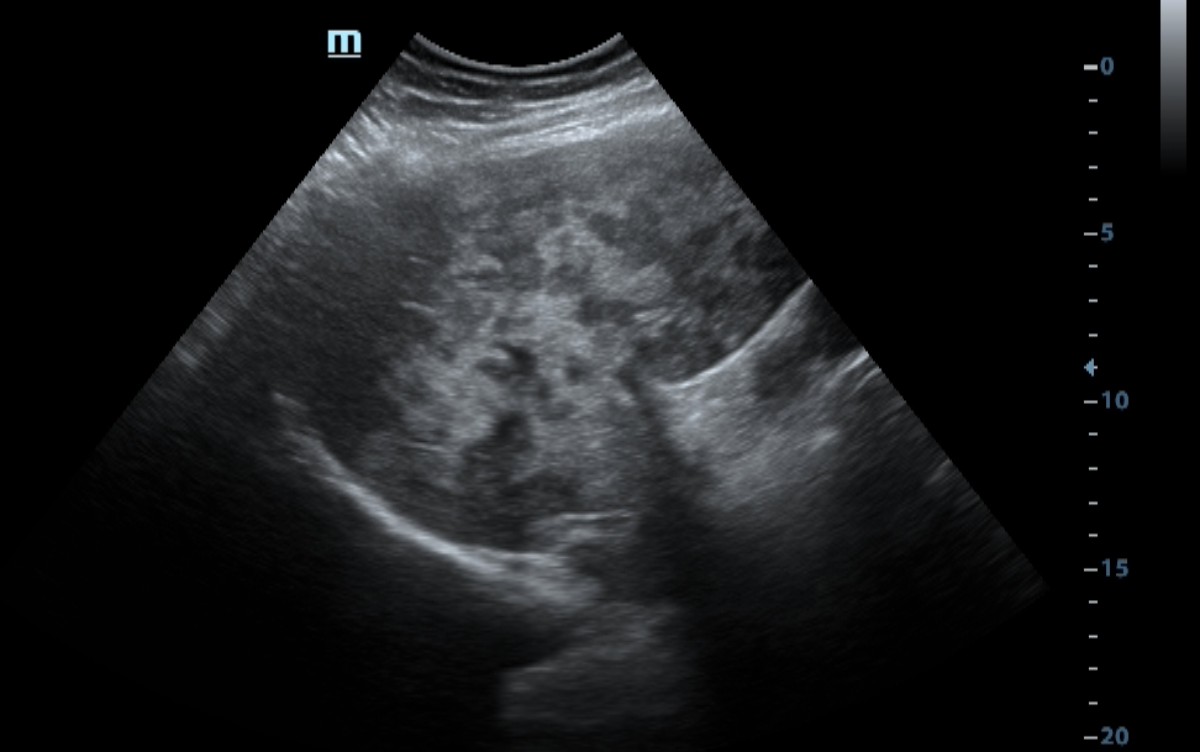
Multiple focal liver lesions were considered metastatic based on their characteristic ultrasound appearance and the patient’s history of breast cancer.
Histopathological confirmation of the liver lesions was not available.
Study method
Ultrasound
Clinical information
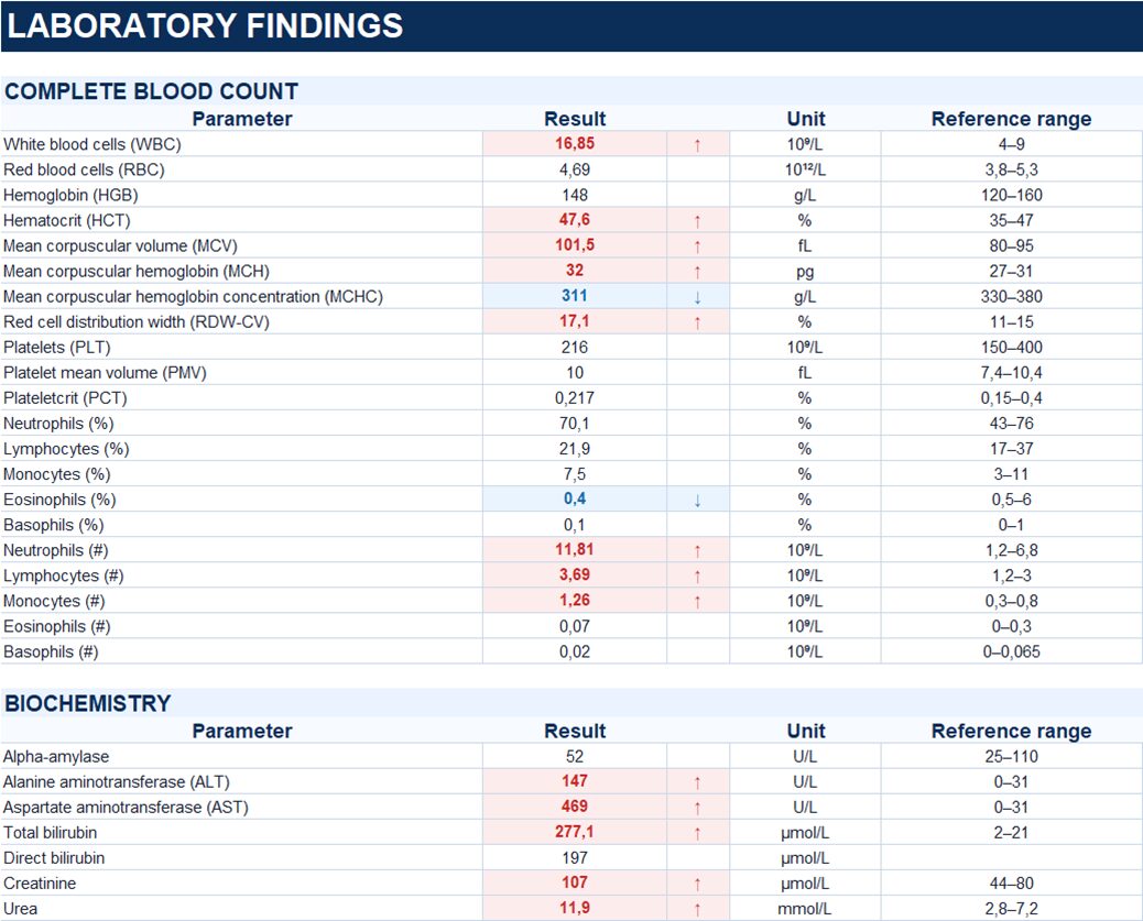
A 58-year-old female was admitted with jaundice of the skin and sclera, dark urine, and epigastric pain. Her medical history was significant for left breast cancer treated with surgery in 2019 followed by chemotherapy.
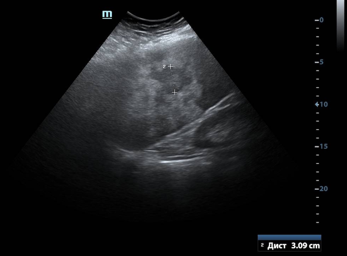
Description of sonographic findings
The liver is enlarged. The hepatic parenchyma is markedly heterogeneous due to multiple focal lesions involving both lobes, some of which coalesce to form irregular conglomerates measuring up to 30 mm. The hepatic vascular architecture is distorted. The portal venous system is not dilated.
The gallbladder is not enlarged, and its wall is not thickened. No gallstones are identified.
The intrahepatic and extrahepatic bile ducts are not dilated.
Conclusion of sonographic findings
Ultrasound findings of hepatomegaly and multiple focal lesions involving both hepatic lobes, most likely metastatic in nature. No signs of biliary dilatation are identified.
Final diagnosis
Parenchymal jaundice. History of left breast cancer. Disease progression with probable metastatic involvement of the liver.
Pitfall
Marked jaundice is not always caused by mechanical biliary obstruction.
In this case, despite marked hyperbilirubinemia, the intrahepatic bile ducts and common bile duct were not dilated. At the same time, hepatomegaly and multiple focal lesions involving both hepatic lobes were identified in a patient with a history of breast cancer.
The absence of biliary dilatation reduces the likelihood of mechanical obstruction of the major bile ducts and warrants consideration of intrahepatic causes of jaundice. In this case, the combination of extensive metastatic liver involvement and the absence of bile duct dilatation supported a predominantly intrahepatic mechanism of jaundice.