
The diagnosis was confirmed by concordant clinical and ultrasound findings.
The diagnosis was confirmed intraoperatively.
The diagnosis was confirmed by histopathological examination of the surgical specimen.
Study method
Ultrasound
Clinical information
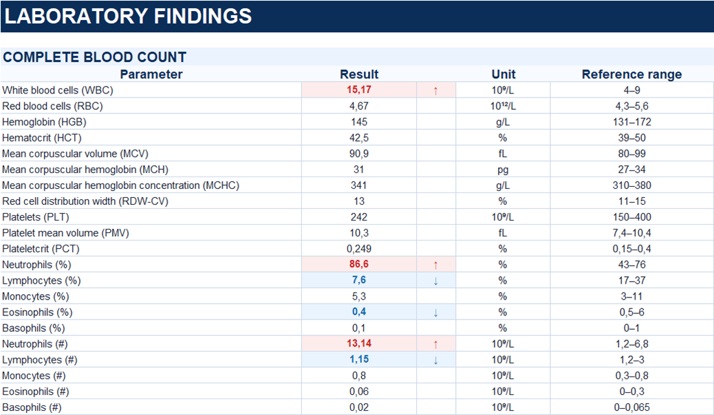
A 46-year-old male presented with right lower quadrant abdominal pain that had started approximately one day earlier and progressively worsened. He denied nausea, vomiting, changes in bowel habits, or urinary symptoms. On admission, his body temperature was 37.2 °C.
Description of sonographic findings
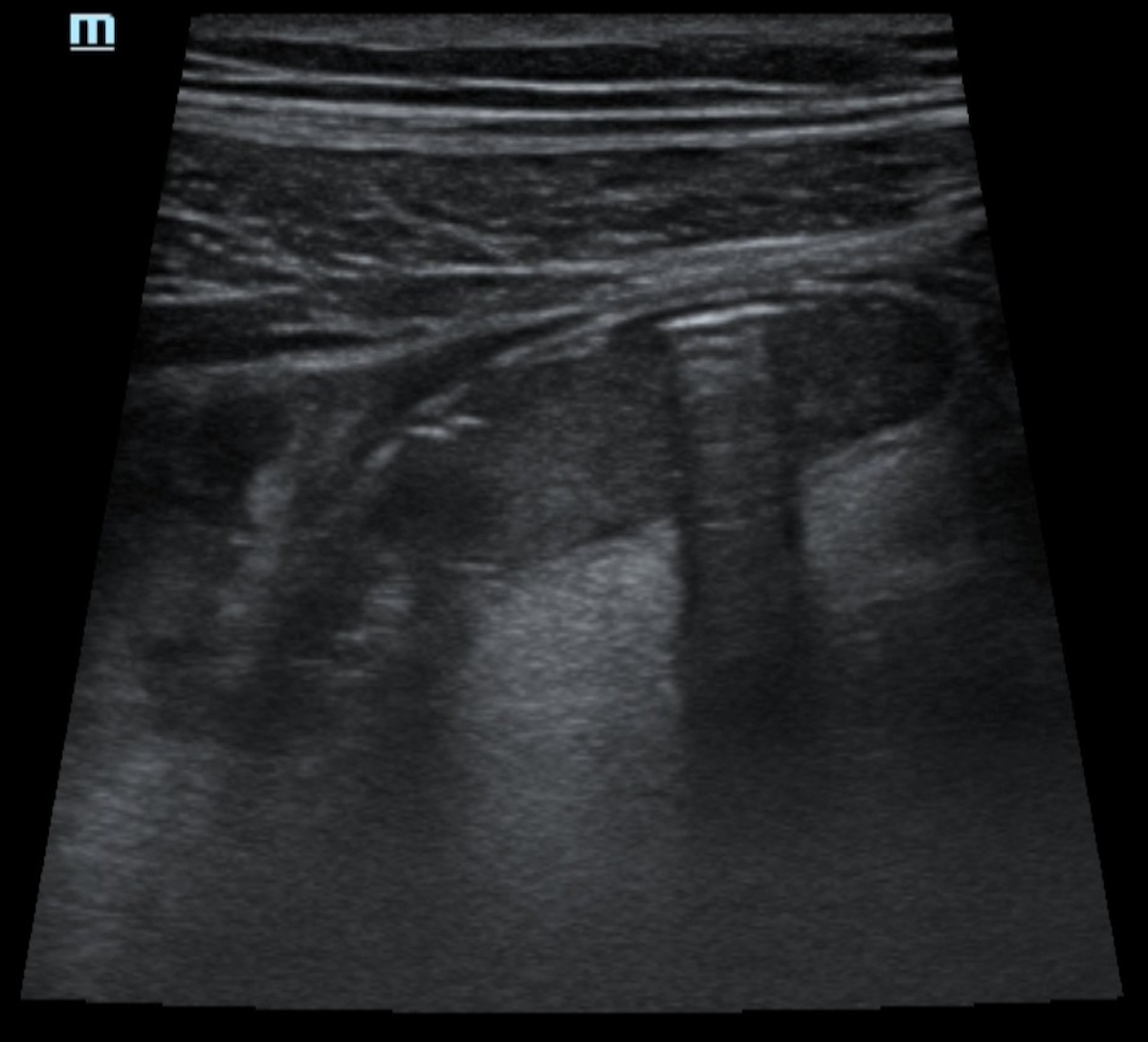
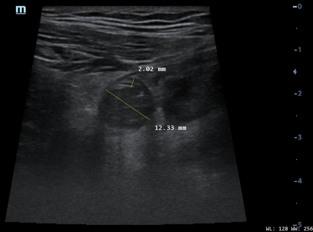
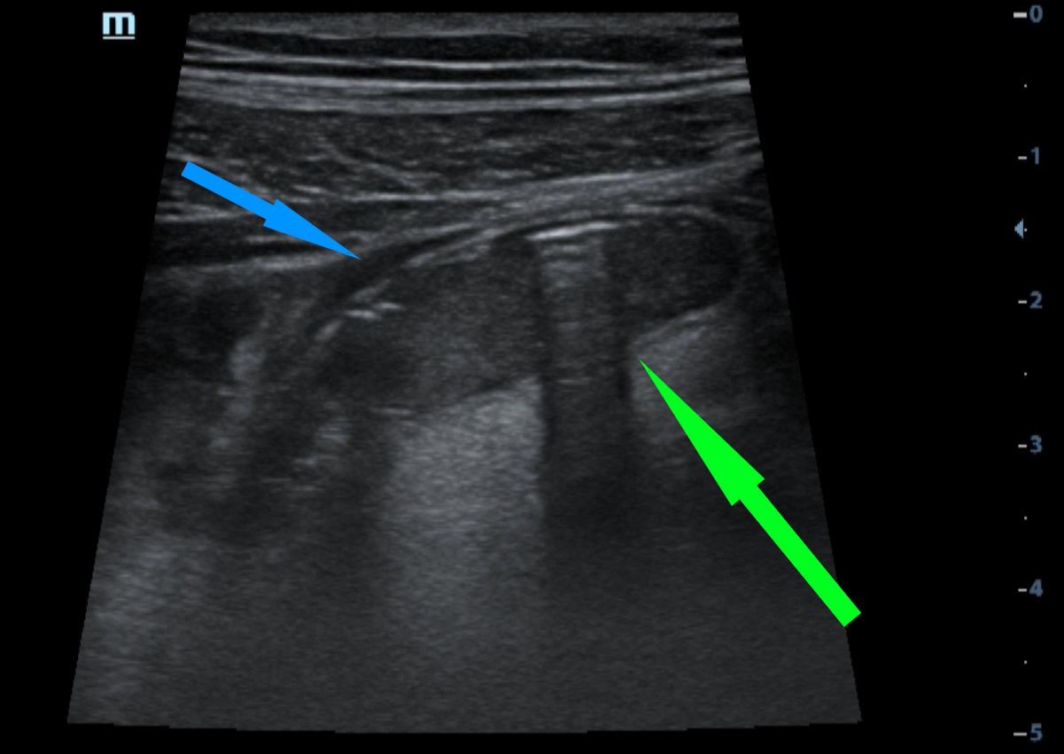
In the right lower quadrant, a blind-ending tubular structure corresponding to the appendix is visualized. The appendix is aperistaltic and non-compressible with transducer pressure.
The outer diameter of the appendix is increased to 12.3 mm; the visualized segment measures approximately 48 mm in length. The lumen is distended and contains heterogeneous hypoechoic material. The wall measures up to 2 mm in thickness, is hypoechoic, and retains its normal layered appearance. A target sign is seen on transverse imaging.
Color Doppler imaging demonstrates increased vascularity of the appendiceal wall.
The periappendiceal fat is moderately hyperechoic and remains compressible. A small amount of free fluid is present around the appendix.
Conclusion of sonographic findings
Ultrasound findings consistent with acute appendicitis.
Treatment
Diagnostic laparoscopy followed by appendectomy was performed.
Intraoperatively, the appendix was thickened, hyperemic, and rigid, with phlegmonous changes. A moderate amount of serous fluid was present in the pelvis.
Verification Method
Histopathology
Histopathological examination demonstrated diffuse, marked neutrophilic infiltration involving all layers of the appendiceal wall, with focal neutrophilic infiltration of the adjacent adipose tissue.
Conclusion: phlegmonous appendicitis, mesenteriolitis, and localized serofibrinous peritonitis.
Final diagnosis
Phlegmonous appendicitis with localized serofibrinous peritonitis.
Pitfall
The severity of individual ultrasound findings may be subtle.
Acute appendicitis does not necessarily present with the full spectrum of classic ultrasound findings. In this case, despite marked enlargement of the appendix to 12.3 mm, the appendiceal wall was not thickened and retained its layered appearance, while the inflammatory changes in the periappendiceal fat were relatively mild.
The diagnosis was based on the combination of findings: a dilated and non-compressible appendix, distension of its lumen, decreased echogenicity and increased vascularity of the appendiceal wall, and a small amount of periappendiceal free fluid.
The absence of marked wall thickening or pronounced inflammatory changes in the surrounding fat does not exclude acute appendicitis. Ultrasound findings should always be interpreted in combination rather than relying on any single feature.