
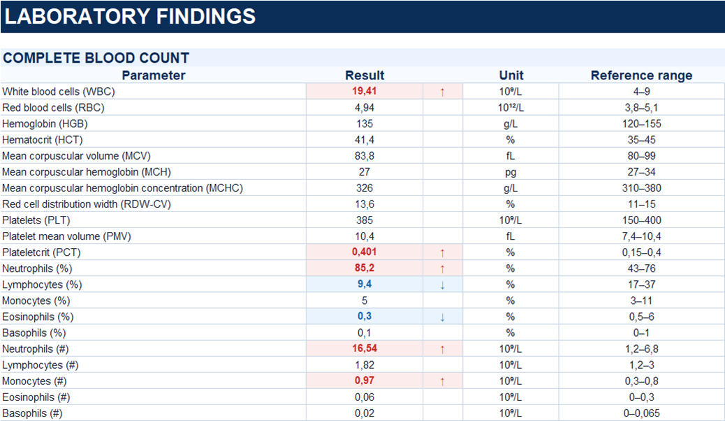
The diagnosis was supported by concordant clinical and ultrasound findings.
The diagnosis was confirmed intraoperatively.
The diagnosis was confirmed by histopathological examination of the surgical specimen.
Study method
Ultrasound
Clinical information
A 31-year-old female presented with discomfort in the right iliac fossa that had developed while she was otherwise well. Due to persistent symptoms, she was admitted to the hospital.
Description of sonographic findings
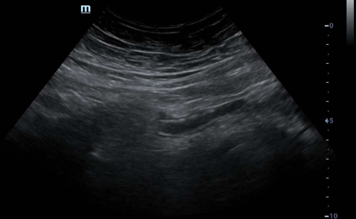
In the right iliac fossa, a non-peristaltic, non-compressible, blind-ending tubular structure corresponding to the appendix is visualized.
The outer diameter of the appendix is increased to 9 mm, with a visualized length of up to 78 mm. The lumen is distended and contains fluid. The wall is thickened to 3 mm and hypoechoic, with preserved mural stratification producing the target sign on transverse imaging.
The periappendiceal fat is moderately hyperechoic. A small amount of periappendiceal free fluid is present, measuring up to 7 mm in depth.
Conclusion of sonographic findings
Ultrasound findings consistent with acute appendicitis. Small amount of periappendiceal free fluid.
Treatment
Diagnostic laparoscopy followed by appendectomy was performed.
Intraoperatively, the appendix was tense and rigid, with marked vascular congestion. A small amount of serous fluid was present in the pelvis. No other pathological changes were identified in the abdominal cavity.
Verification Method
Histopathological examination
Microscopic examination revealed superficial mucosal necrosis and moderate diffuse neutrophilic infiltration extending through the entire thickness of the appendiceal wall and into the adjacent adipose tissue. Vascular congestion and hemorrhage were also present.
Conclusion: phlegmonous ulcerative appendicitis, mesenteriolitis, and periappendicitis.
Final diagnosis
Phlegmonous ulcerative appendicitis.