

The diagnosis was confirmed by concordant clinical, ultrasound, and CT findings.
Purulent material was obtained during percutaneous drainage of the abscess.
Study method
Ultrasound, CT
Clinical information
The patient reported a one-month history of pain in the left iliac fossa. At presentation, she also complained of lower abdominal pain and urinary frequency. She denied nausea, vomiting, and fever. Due to persistent symptoms, she was admitted to the hospital for further evaluation.
Description of sonographic findings
In the left pelvis, within the pericolic fat adjacent to the sigmoid colon and the urinary bladder dome, there is a heterogeneous fluid collection measuring up to 38 × 24 mm, containing hyperechoic foci consistent with gas.
The surrounding fat demonstrates increased echogenicity, consistent with perifocal inflammatory changes. The urinary bladder wall is mildly thickened at the site adjacent to the collection. Multiple diverticular outpouchings are also identified along the sigmoid colon.
Conclusion of sonographic findings
Sonographic findings of a gas-containing pelvic abscess with surrounding inflammatory changes. Mild reactive thickening of the adjacent urinary bladder wall. Multiple sigmoid diverticula.


Portal venous phase, axial plane.
Portal venous phase, sagittal MPR
Description of radiographic findings
In the left pelvis, there is a well-defined multiloculated fluid collection measuring approximately 70 × 20 mm, containing gas bubbles and demonstrating peripheral rim enhancement.
The collection is inseparable from the thickened sigmoid colon, which contains multiple diverticula. It also abuts the thickened urinary bladder wall and the anterior abdominal wall.
Conclusion of radiographic findings
CT findings of acute sigmoid diverticulitis complicated by a pericolic pelvic abscess (Hinchey II).
Treatment
Ultrasound-guided percutaneous drainage of the pelvic abscess was performed with placement of a 10 Fr pigtail catheter. Approximately 20 mL of purulent material was aspirated. Follow-up ultrasound demonstrated a decrease in the size of the abscess cavity, with the drainage catheter positioned within it.
Final diagnosis
Diverticular disease. Acute sigmoid diverticulitis complicated by a pericolic pelvic abscess (Hinchey II).
Pitfall
Gas may hinder sonographic assessment of an abscess
Gas within an abscess appears as hyperechoic foci associated with reverberation artifacts and dirty acoustic shadowing. These artifacts may partially obscure the fluid component and abscess wall, making the collection appear less well-defined on ultrasound than on CT.
The absence of a clearly visualized capsule does not exclude an abscess. A combination of a fluid collection, intralesional gas, and inflammatory changes in the surrounding fat should raise suspicion for an infected collection.
Additional information
Clinical and diagnostic reasoning
- The sigmoid diverticula provide the underlying structural substrate for the development of acute diverticulitis. Extension of the inflammatory process beyond the bowel wall resulted in the formation of a pericolic abscess with marked inflammatory changes in the surrounding fat.
- The abscess is located immediately adjacent to the urinary bladder. Mild thickening of the adjacent bladder wall is likely reactive and provides an anatomical explanation for the patient’s urinary frequency, alongside the left iliac fossa pain typical of sigmoid diverticulitis.
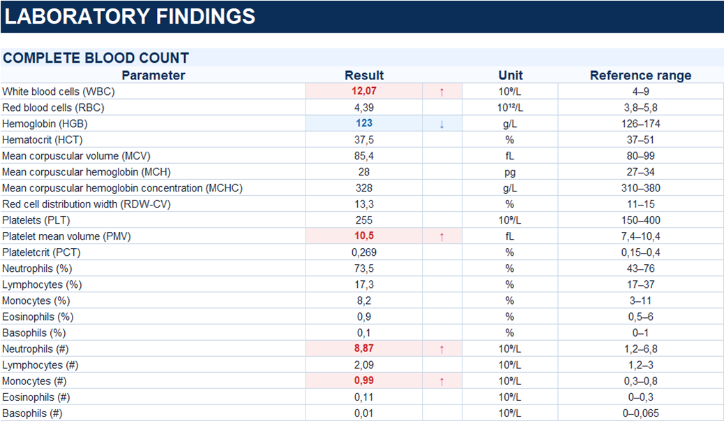
- Mild leukocytosis with absolute neutrophilia further supports the presence of an active inflammatory process. Thus, the clinical symptoms, laboratory abnormalities, and imaging findings represent different manifestations of the same pathological process: acute sigmoid diverticulitis → pericolic abscess formation → surrounding inflammation with reactive changes in the adjacent urinary bladder wall.