

The diagnosis was confirmed by concordant ultrasound and CT findings.
The findings on both imaging modalities were consistent with acute diverticulitis.
Study method
Ultrasound, CT
Clinical information
Female patient, 53 years old, presented to the emergency department with pain in the left lateral abdomen. The pain was associated with changes in bowel habits. The patient reported no nausea or vomiting.
Complete blood count: moderate leukocytosis (13 × 10⁹/L) with neutrophilia (83.2%).


Description of sonographic findings
In the left lateral abdomen, a focally thickened segment of the descending colon is visualized. Along the colonic wall, a diverticulum measuring up to 10 mm is identified, containing a hyperechoic component with posterior acoustic shadowing, likely due to gas and/or dense fecal material.
The adjacent colonic wall measures 9–12 mm in thickness. The surrounding pericolic fat demonstrates increased echogenicity, consistent with inflammatory changes. No free fluid or organized fluid collections are identified.
Conclusion of sonographic findings
Ultrasound findings are consistent with diverticulitis of the descending colon.
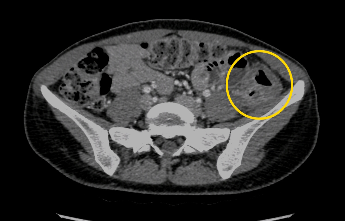
Порто-венозная фаза
Description of radiographic findings
In the left lateral abdomen, diverticula of the descending colon are identified, with local circumferential wall thickening. The surrounding pericolic fat demonstrates inflammatory infiltration.
No free intraperitoneal gas or organized fluid collections are identified.
Conclusion of radiographic findings
CT findings are consistent with diverticulitis of the descending colon without signs of a complicated abscess.
Treatment
Conservative treatment was initiated, including antibiotic therapy and symptomatic management, with subsequent clinical improvement.
As no urgent surgical intervention was indicated, the patient was discharged in satisfactory condition after stabilization.
Final diagnosis
Diverticulitis of the descending colon. Hinchey Ia.
Pitfall
При локальной боли в левом фланке дивертикулит нисходящей ободочной кишки может быть пропущен, если исследование ограничивается оценкой паренхиматозных органов и почек.
Ключевым является целенаправленное исследование кишечника в зоне максимальной болезненности. Выявление локального утолщения стенки кишки, воспалённого дивертикула и гиперэхогенной изменённой окружающей клетчатки позволяет предположить диагноз уже при УЗИ.