
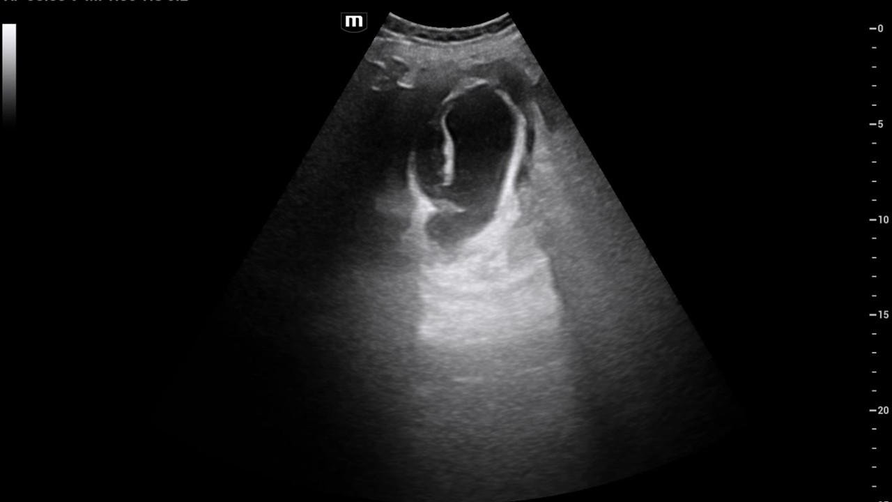
The diagnosis was confirmed by concordant ultrasound and CT findings.
The diagnosis was also confirmed intraoperatively.
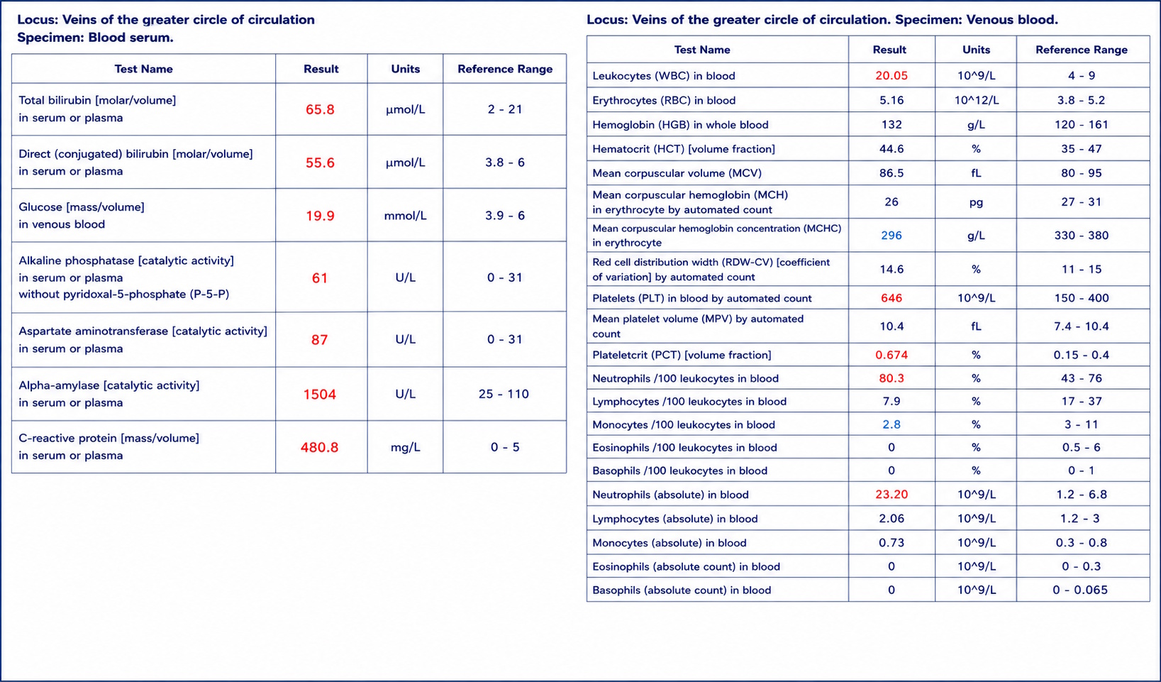
Predominantly direct hyperbilirubinemia and a moderate elevation of liver transaminases were present.
A markedly elevated serum α-amylase level was also noted; however, CT demonstrated no evidence of acute pancreatitis.
Significant hyperglycemia was also present.
Study method
Ultrasound, CT
Clinical information
A 72-year-old woman presented with a two-week history of upper abdominal pain, initially accompanied by nausea and repeated vomiting. She did not seek medical attention. Due to persistent abdominal pain and fever up to 39°C, she was transported by ambulance to the emergency department.

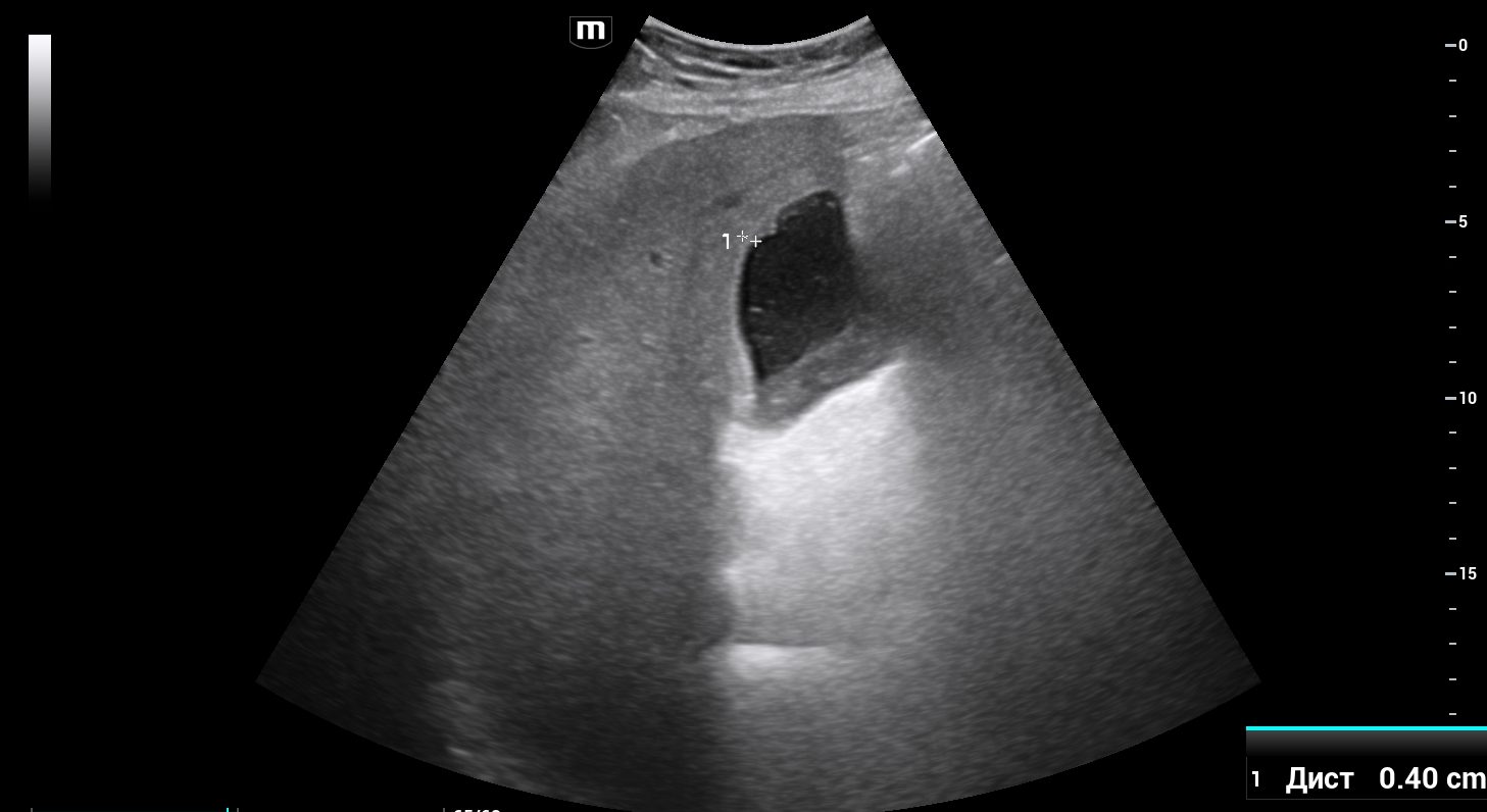
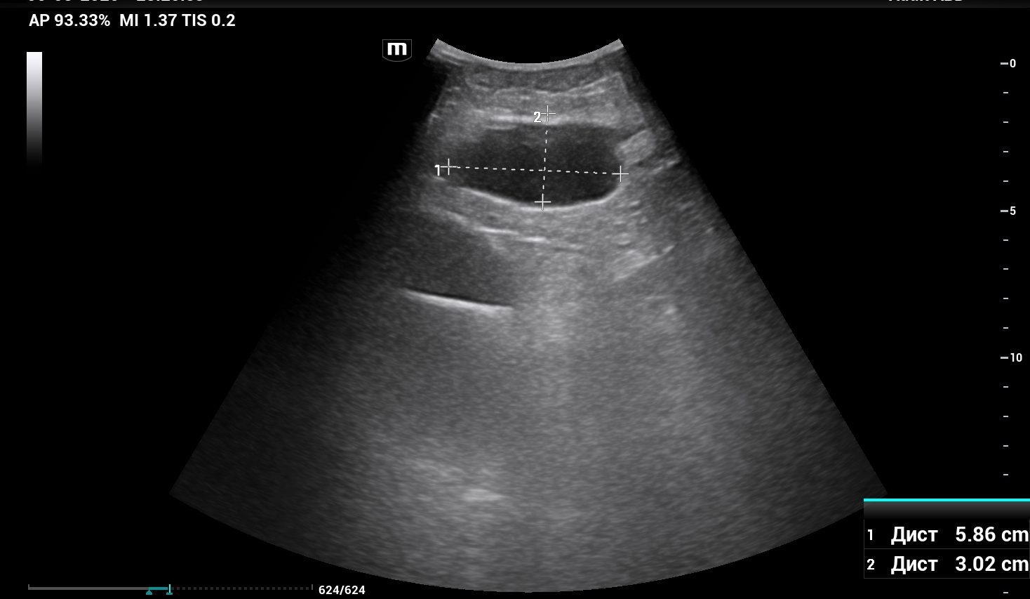
Description of sonographic findings
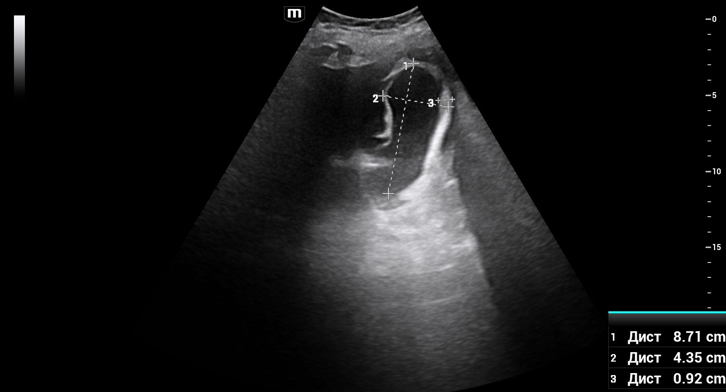
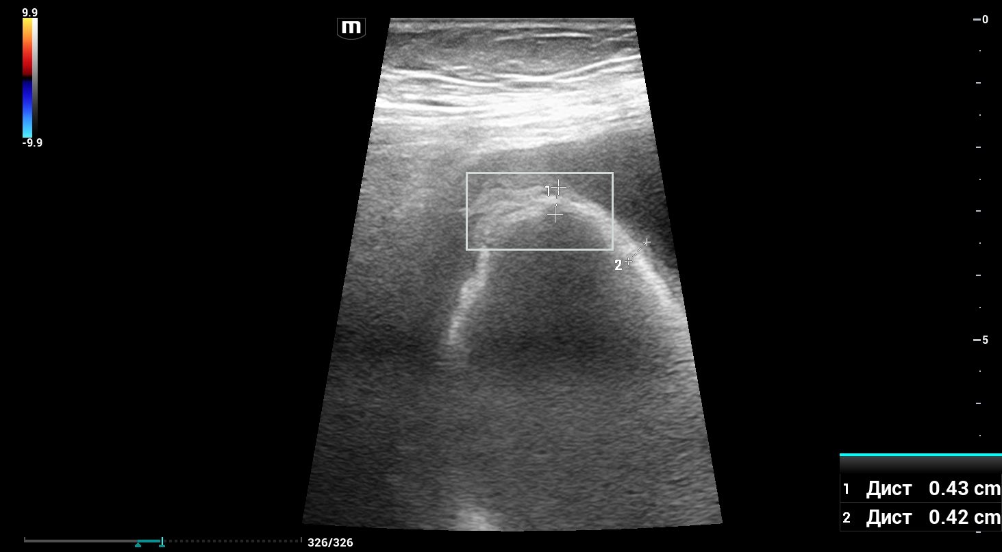
The gallbladder is deformed and measures 81 × 34 mm. The gallbladder wall is irregularly thickened up to 5 mm, demonstrates preserved layering, and contains focal areas of wall thinning and contour disruption. A heterogeneous pericholecystic fluid collection measuring up to 24 mm in thickness is identified, consistent with a pericholecystic abscess.
A well-defined subhepatic fluid collection beneath the left hepatic lobe demonstrates thick walls and heterogeneous internal contents, suspicious for an abscess.
Within the epigastrium, an additional encapsulated fluid collection measuring 58 × 30 mm is identified. It abuts the parietal peritoneum anteriorly and is bordered inferiorly by the greater omentum, also suspicious for an abscess.
Conclusion of sonographic findings
Ultrasound findings are consistent with:
Acute cholecystitis complicated by a pericholecystic abscess.
Additional encapsulated heterogeneous fluid collections in the subhepatic and epigastric regions, most consistent with abscesses.
Порто-венозная фаза
Description of radiographic findings
The gallbladder is enlarged, measuring approximately 15 × 7 cm.The gallbladder wall is not significantly thickened; however, inflammatory fat stranding surrounds the gallbladder. The lumen contains heterogeneous material without convincing visualization of radiopaque gallstones.
Two irregular fluid collections measuring 6 × 5 cm and 6 × 1.5 cm are identified within the subhepatic region, extending into hepatic segment III.
An additional multiloculated encapsulated fluid collection measuring approximately 11 × 7.5 cm is located anterior to the liver along the falciform ligament, extending into the anterior abdominal wall musculature.
Conclusion of radiographic findings
CT findings are consistent with destructive acute cholecystitis complicated by multiple abscesses involving: the liver, the subhepatic space, the anterior abdominal wall.
Treatment
Given the prolonged disease course and the presence of well-formed localized abscesses, ultrasound-guided percutaneous treatment was performed.
Three procedures were completed during a single session:
1) Percutaneous transhepatic cholecystostomy, yielding approximately 230 mL of thick purulent material mixed with bile.
2) Percutaneous drainage of the abscess adjacent to hepatic segment V, evacuating approximately 200 mL of thick purulent material mixed with bile.
3) Transhepatic drainage of the left subhepatic abscess, evacuating approximately 200 mL of thick purulent material mixed with bile.
Separate drainage catheters were placed into all three collections under ultrasound guidance.
In total, approximately 630 mL of purulent bile-stained material was evacuated.
Final diagnosis
Cholelithiasis. Acute destructive cholecystitis complicated by perforation into the peritoneal cavity with formation of multiple abscesses involving: hepatic segment V, the left subhepatic space, the anterior abdominal wall.
Pitfall
In patients with prolonged destructive cholecystitis, secondary abscesses may become the dominant imaging finding and obscure the primary source of infection.
The presence of multiple fluid collections within the liver, subhepatic space, and anterior abdominal wall should prompt careful evaluation of their anatomical continuity and a targeted search for the underlying infectious source. In this case, the key diagnostic clues were the severely abnormal gallbladder and the recovery of purulent bile-stained material from all three drained collections.

Автор
Исаев Егор Алексеевич
Врач УЗИ, рентгенолог
Автор образовательного проекта с клиническими случаями по ультразвуковой и лучевой диагностике.