
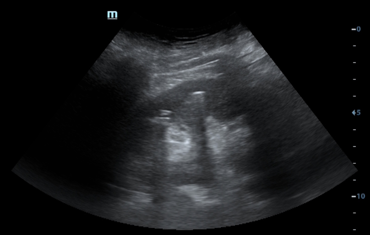
The diagnosis was confirmed by concordant ultrasound and CT findings.
The diagnosis was also confirmed intraoperatively.
Study method
Ultrasound, CT
Clinical information
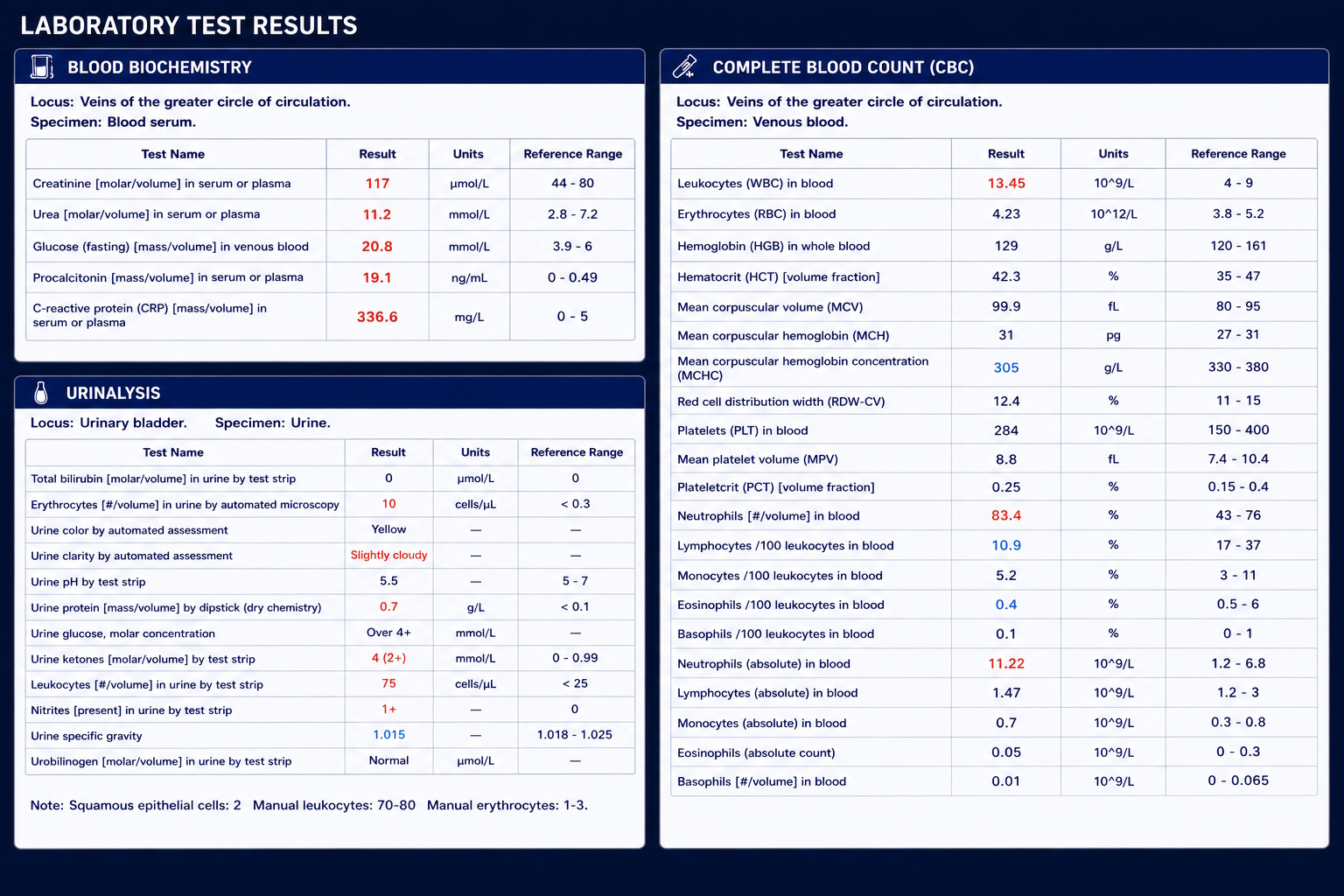
A 69-year-old woman presented with a one-week history of progressively worsening generalized weakness, drowsiness, excessive thirst, dry mouth, polyuria, and loss of appetite. On the day of admission, due to marked clinical deterioration, she was transported by ambulance to the emergency department. Her medical history was significant for type 2 diabetes mellitus and arterial hypertension.

Description of sonographic findings
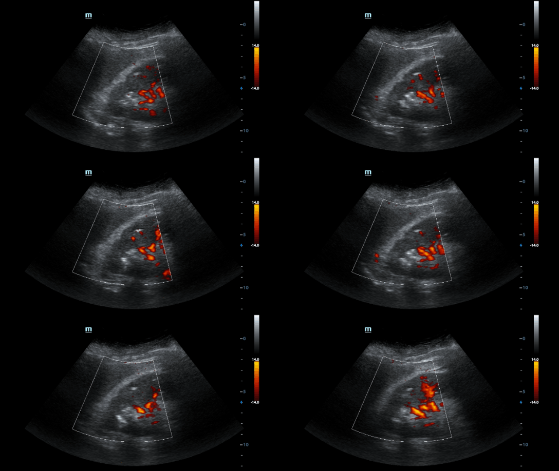
The right kidney is normal in size (109 × 60 mm) and normally positioned. Renal parenchymal thickness measures 18 mm and demonstrates diffusely heterogeneous echotexture. Within the upper pole, multiple punctate hyperechoic foci with posterior comet-tail artifacts are identified, a highly specific finding for intraparenchymal gas. Color Doppler imaging demonstrates reduced parenchymal perfusion within the upper pole. The collecting system is not dilated. No renal calculi producing posterior acoustic shadowing are identified. The ureter is not dilated.
Conclusion of sonographic findings
Ultrasound findings are highly specific for emphysematous pyelonephritis of the right kidney, demonstrated by intraparenchymal gas and reduced renal perfusion.
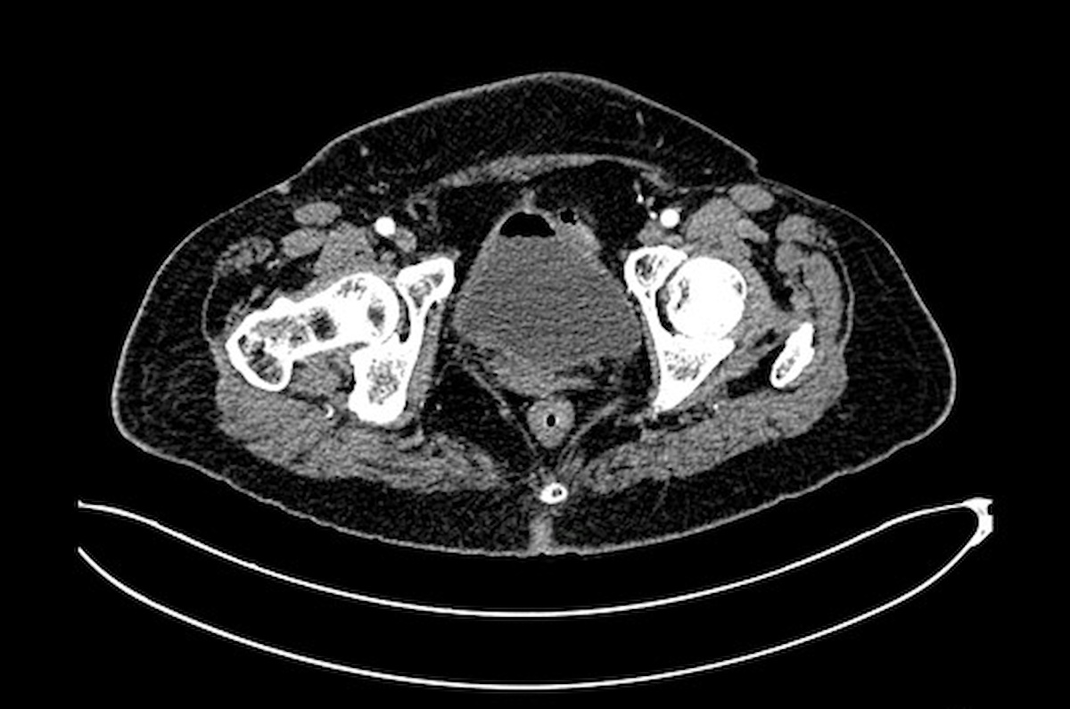
Description of radiographic findings
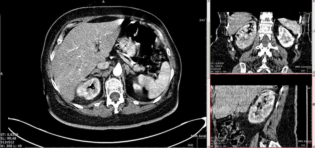
Multiple rounded and wedge-shaped hypoenhancing areas are present throughout the right renal parenchyma with loss of normal corticomedullary differentiation. Within these regions, multiple small low-attenuation foci measuring up to 6 mm (approximately +25 HU) are identified, corresponding to areas of suppurative parenchymal destruction. Numerous collections of gas are present within the renal pyramids and the collecting system. Diffuse circumferential thickening of the walls of the renal collecting system and the entire right ureter is observed. Moderate inflammatory stranding of the right perinephric fat is present. The urinary bladder wall is irregularly thickened up to 7 mm, and gas is identified within the bladder lumen.
Conclusion of radiographic findings
CT findings are consistent with right-sided emphysematous pyelonephritis with multiple foci of suppurative renal parenchymal destruction. Associated inflammatory thickening of the collecting system, right ureter, and urinary bladder wall is present, along with intravesical gas.
Treatment
Due to the severe course of emphysematous pyelonephritis and extensive destructive involvement of the right kidney, an emergency right nephrectomy was performed.
Intraoperative Findings:
– Enlarged right kidney with a soft, friable consistency.
– Numerous cortical microabscesses (apostemata) distributed throughout the kidney, some of which were confluent.
– Gerota’s fascia demonstrated extensive fibrinous-necrotic changes.
– Severe destructive renal infection consistent with emphysematous pyelonephritis.
Given the irreversible nature of the renal destruction, a right nephrectomy was performed.
Because of diffuse retroperitoneal bleeding, temporary packing of the surgical wound was required.
Following hemodynamic stabilization, a planned second-look procedure was performed with removal of the packing material and definitive drainage of the retroperitoneal space.
Final diagnosis
Acute right-sided emphysematous pyelonephritis. Sepsis. Type 2 diabetes mellitus with diabetic ketosis.
Pitfall
Gas within the kidney may be mistakenly interpreted as renal calculi, bowel reverberation artifacts, or calcifications within the collecting system.
Key points to consider:
– multiple bright hyperechoic foci;
– prominent reverberation (“dirty shadowing”) artifacts;
– absence of the clean posterior acoustic shadow typical of calculi;
– correlation with the patient’s severe clinical condition.

Author
Egor Isaev
Ultrasound physician, Radiologist
Author of an educational project featuring clinical cases in ultrasound and diagnostic imaging.